German Research Foundation supports MHH Department of Nuclear Medicine with around 900,000 euros for the expansion of molecular imaging

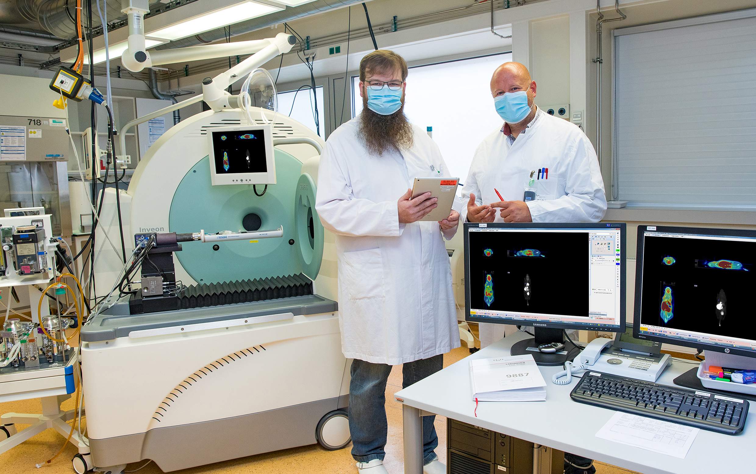
Dr. Jens Bankstahl (left) and Professor Dr. Tobias Ross next to a combination device for preclinical positron emission tomography/computed tomography (PET/CT) in the Department of Nuclear Medicine ; Copyright: Karin Kaiser / MHH
24.11.2021
In order to detect and research diseases, it is important to look inside the body. For this purpose, there are various imaging methods – from ultrasound examinations to X-rays and computer tomography. Molecular imaging provides a particularly precise insight, showing biological processes and organ functions "live" and can thus reveal the course of a disease or the effectiveness of a treatment at an early stage. In this regard, the Department of Nuclear Medicine at Hannover Medical School (MHH) has been offering technology and expertise to research groups inside and outside the MHH to support their scientific work since 2012. With the new funding as a "core facility" on the topic of "Standardised Preclinical Molecular Imaging with Flexible Radiopharmaceuticals", the department wants to improve the offer even further in future, expand the infrastructure and provide a kind of toolbox for the various needs relating to the scientific questions. The German Research Foundation (DFG) is funding the project with about 900,000 euros over five years. "This is a project that is unique in Germany in this form," emphasises Professor Dr. Frank Bengel, Director of the Department of Nuclear Medicine and Dean of Research at MHH.
Tiny sniffing substances find cells according to the lock-and-key principle
"Molecular imaging makes biological mechanisms and organ functions in the living organism visible and is therefore enormously important for medical research," explains Professor Dr. Tobias Ross, Head of the Department of Radiopharmaceutical Chemistry. This is achieved with the help of so-called radiotracers or radiopharmaceuticals, which are administered into the body via injection. The tiny tracers are weakly radioactive for only a short time and can be visualised, for example, by high-resolution positron emission tomography (PET). If, for instance, certain sugar molecules are radioactively labelled, they detect regions in the body that consume an unusually large amount of energy. These include tumours, for example. The radiotracers can also be selected in such a way that they attach themselves specifically to the surface of certain cells according to the lock-and-key principle and thus mark possible therapeutic targets in the body.
With molecular imaging to personalised medicine
"With the help of DFG funding, we want to standardise our methods and make them accessible to all research groups as a platform technology," explains Dr. Jens Bankstahl, Head of the Clinical Department Preclinical Molecular Imaging. The Core Facility wants to offer flexible and innovative services as well as model solutions for regularly recurring questions. Another goal is to develop and test new radiotracers that can be used in the clinic for diagnostics and treatment later. "Over the course of time, we have already transferred some radiopharmaceuticals and imaging methods into practical application," Professor Ross emphasises. Now, he says, the aim is to expand the spectrum and find new, precisely fitting therapeutic approaches for infectious diseases, cancer or cardiovascular diseases. "This is the decisive step towards personalised medicine, in which each patient is treated individually according to the respective clinical picture."
SERVICE:
For further information, please contact Professor Dr. Tobias Ross, ross.tobias@mh-hannover.de, telephone (0511) 532-5895 or Dr. Jens Bankstahl, bankstahl.jens@mh-hannover.de, telephone (0511) 532-3504.