AG Iorga

Contraction-relaxation function and mechano-chemical coupling of myofibrils
Investigations of human, human pluripotent stem cell-derived cardiomyocytes and animal contractile models in non-pathologic and pathologic conditions
Forschungsschwerpunkt
Contraction-relaxation function and mechano-chemical coupling of myofibrils.
The main function of cardiomyocytes (CMs) to generate force and shorten their length occurs when the subcellular myofibrils contract due to multiple interactions between ATPase-driven myosin motors and actin filaments. Myofibrils consist in many sarcomeres arranged in series driving directly the contraction-relaxation events of CMs upon cyclical variation of the intracellular Ca2+ concentration. Therefore, isolated myofibrils represent a contractile model used to understand sarcomeric protein-related processes that determine contractile function of CMs in the absence of Ca2+ handling systems and of upstream signaling.
Myofibrils can be investigated using fast kinetic micromechanical and chemical techniques because they are thin and in rapid diffusional equilibrium with their surrounding environment. We have established a micromechanical setup that uses an atomic force cantilever as a nN-sensitive force sensor. This setup allows rapid changes of the solutions to which myofibrils are exposed, and the force kinetic parameters of myofibrillar activation and relaxation at different Ca2+ concentrations can be analyzed with high time resolution. Different established biochemical methods can assess the steady-state or transient ATPase activity of the sarcomeric myosin motor using mechanical unloaded, native myofibrils or using myofibrils prevented from shortening by chemical cross-linking few of their myosin heads to the actin filaments. These investigations can allow correlating the biochemical events to the mechanical events during cross-bridge cycling based on isoform composition of sarcomeric proteins.
Subcellular myofibrils can be obtained from human (e.g., biopsies), human-derived and non-human cardiac and skeletal small muscle samples (e.g., mouse, rat, rabbit, zebrafish and hESC-/hiPSC-CMs).
In vitro-differentiated cardiomyocytes.
Human pluripotent stem cell-derived cardiomyocytes (hPSC-CMs) hold great potential for the treatment of cardiovascular diseases by cell transplantation or engineered cardiac tissue, for assessing efficiency and toxicity of pharmacological compounds, or to be used as cellular disease models in vitro. hPSC-CMs exhibit a series of immature features compared to ventricular CMs of an adult human heart. Therefore, characterization of hPSC-CMs at different hierarchically interrelated levels (molecular, subcellular, cellular and multicellular levels) and understanding how extracellular environment and intracellular factors may affect subcellular myofibrils and cell maturation of hPSC-CMs, in pathologic and non-pathologic conditions, represents important research objectives to us.
Our research interests are related to:
- understanding and identification of biophysical factors that could improve the functional maturation of hPSC-CMs in vitro toward a more ventricular-like phenotype;
- defining the maturation stage of hPSC-CMs at different hierarchically interrelated levels (molecular, subcellular, cellular and engineered tissue levels) with the purpose to further use hPSC-CMs as contractile model for different cardiomyopathies;
- understanding the functional primary consequences of missense mutations (e.g., cardiomyopathy-linked mutations) occurring in sarcomeric proteins (e.g., in myosin motor) using the hPSC-CMs model at different maturation stages in comparative studies with adult human myocardial samples;
- myofibrillar phenotypization involving assessments with multiple micromechanical and biochemical methods for studies of development, regeneration and inherited diseases.
Current research topics:
- Application of biophysical stimuli to promote myofibrillar maturation of human pluripotent stem cell-derived cardiomyocytes
- Investigations on the contractile and biochemical function of myofibrils isolated from myocardium and cardiomyocytes in cell culture in non-pathologic and pathologic conditions
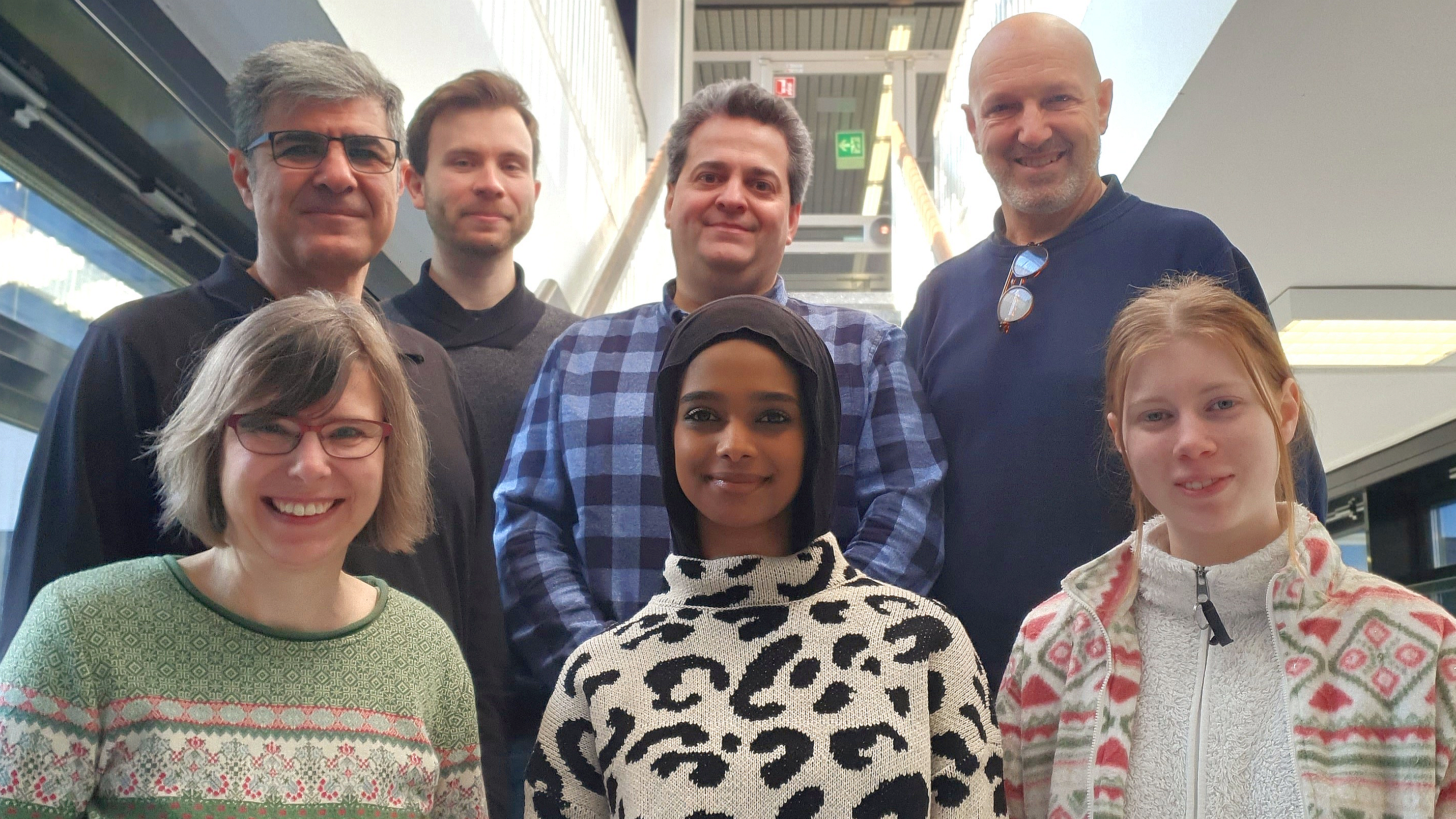
Group members:
Shared personal:
- Dr. rer. nat Meißner Joachim (molecular and cell biology)
- Birgit Piep (proteins analysis)
- Dipl.-Ing. (FH) Faramarz Matinmehr (Cardiomyozyten)
- Dr. rer. nat. Ante Radocaj (statistik und modelling)
- Alexander Lingk (mechanics)
- Torsten Beier (optics and optoelectronics)
- Uwe Krumm (electronics)
Methods:
- Micromechanical investigation of contractile function of myofibrils
- Biochemical analysis of sarcomeric proteins
read more
Related (selected) publications:
- Oscillatory work and the step that generates force in single myofibrils from rabbit psoas, M. Kawai and B. Iorga, Pflugers Arch - Eur J Physiol., 2024, April 1, https://doi.org/10.1007/s00424-024-02935, Pubmed
- Myosin expression and contractile function are altered by replating stem cell–derived cardiomyocytes, Osten, F., Weber, N., Wendland, M., Holler, T., Piep, B., Kröhn, S., Teske, J., Bodenschatz, A.K., Devadas, S.B., Menge, K.S., Chatterjee, S., Schwanke, K., Kosanke, M., Montag, J., Thum, T., Zweigerdt, R., Kraft, T., Iorga, B., and Meissner, J.D. (2023), J Gen Physiol (2023) 155 (11): e202313377, https://doi.org/10.1085/jgp.202313377, Online ahead of print, PubMed
- M. Kawai, R. Stehle, G. Pfitzer and B. Iorga, Phosphate has dual roles in cross-bridge kinetics in rabbit psoas single myofibrils, J. Gen. Physiol. 2021 Vol. 153 No. 3, DOI: 10.1085/jgp.202012755, PubMed
- Iorga B., Schwanke K., Weber N., Wendland M., Greten S., Piep B., dos Remeidos C.G., Martin U., Zweigerdt R., Kraft T., Brenner B. (2018) “Differences in Contractile Function of Myofibrils within Human Embryonic Stem Cell-Derived Cardiomyocytes vs. Adult Ventricular Myofibrils Are Related to Distinct Sarcomeric Protein Isoforms“, Frontiers in Physiology, 8:1111 DOI:10.3389/fphys.2017.01111 PubMed
- Weber N., Greten S., Wendland M., Schwanke K., Iorga B., Fischer M., Geers-Knörr C., Hegermann J., Wrede C., Fiedler J., Kempf H., Franke A., Piep B., Pfanne A., Thum T., Martin U., Brenner B., Zweigerdt R., Kraft T. (2016) “Stiff matrix induces switch to pure β-cardiac myosin heavy chain expression in human ESC-derived cardiomyocytes”, Basic Research in Cardiology, 111:68 DOI: 10.1007/s00395-016-0587-9 PubMed
- Elhamine F., Iorga B., Krüger M., Hunger M., Eckhardt J., Sreeram N., Bennink G., Brockmeier K., Stehle R., Pfitzer G. (2016) “The postnatal development of right ventricular myofibrillar biomechanics in relation to the sarcomeric protein phenotype in pediatric patients with conotruncal heart defects”, Journal of the American Heart Association, 5:e003699 DOI:10.1161/JAHA.116.003699 PubMed
- Iorga B., Neacsu C.D., Neiss W.F., Wagener R., Paulsson M., Stehle R., Pfitzer G. (2011) “Micromechanical function of myofibrils isolated from skeletal and cardiac muscles of the zebrafish”, The Journal of General Physiology (ISSN: 0022-1295), Vol.137(3):255-270 PubMed
- Lionne C., Iorga B., Candau R., Travers F. (2003) “Why choose myofibrils to study muscle myosin ATPase?”, J. Muscle Res. Cell Motil. (ISSN: 0142-4319), Vol. 24 (2-3):139-148 PubMed
Didactic activities:
- LabCourse for Biomedicine Master students: “Functional Investigations of Myofibrils” (Hannover Biomedical Research School, HBRS)
- LabCourses for Human and Stomatologic students: “Physical and Physiological Basis for Medicine” (Hannover Medical School, MHH)
Collaborations:
- Internal (MHH): Leibniz Research Laboratories for Biotechnology and Artificial Organs (LEBAO), HTTG-Chirurgie, NIFE
- External: University of Köln, University of Bucharest, University of Sydney