Master’s Thesis Position:
Photophysical Characterization of Next-Generation Ca2+ Biosensors for Quantitative Astrocyte Imaging

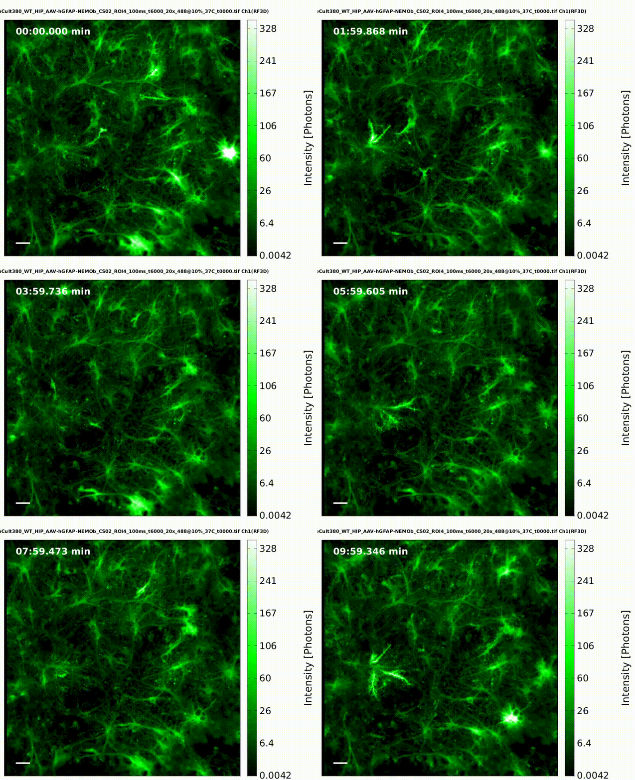
About the Project: Genetically encoded calcium biosensors are transforming quantitative functional imaging of glial cells by enabling precise readouts of intracellular signaling. Our mission is to rigorously characterize newly developed Ca2+ biosensor variants based on NEMO-V5-turboFP650 and to establish their utility for high-fidelity, quantitative imaging in primary cultures of mouse hippocampal astrocytes. You will help define the photophysical and functional performance limits of these probes and assess how they can open new avenues for astrocyte research.
Join Our Dynamic Team: Become an integral member of the Cellular Neurophysiology team at Hannover Medical School, working within cutting-edge microscopy and cell culture facilities. Contribute to a timely and impactful project under the guidance of experienced supervisors. We provide comprehensive mentoring and hands-on training to support your success. Our expertise and infrastructure include:
- Advanced fluorescence microscopy (widefield, confocal, spinning disk; optional FLIM)
- Quantitative spectroscopy and probe characterization
- Primary mouse astrocyte culture and live-cell imaging workflows
- Image processing, quantitative analysis, and scripting (Python/Matlab)
- Access to high-performance computing resources for data analysis
Your Role: As a Master’s thesis candidate, you will:
- Determine key photophysical parameters of NEMO-V5-turboFP650 variants (absorption/emission spectra, extinction coefficient, quantum yield, brightness, photostability, pH sensitivity)
- Quantify Ca2+ sensing performance (dynamic range, affinity/Kd, on/off kinetics, baseline stability) using in vitro titrations and in-cell calibrations
- Establish imaging protocols for quantitative functional Ca2+ mapping in primary mouse hippocampal astrocytes under low-light conditions
- Evaluate sensor performance in live cells (ΔF/F0, signal-to-noise, photobleaching rates, response linearity, compatibility with multi-color imaging)
- Compare against established Ca2+ indicators and assess applications to astrocyte-specific questions (e.g., microdomain activity in fine processes, neurotransmitter-evoked responses)
- Develop robust analysis pipelines for segmentation, event detection, and quantitative metrics; document methods to ensure reproducibility
You Bring:
- Background in biophysics, neuroscience, biomedical engineering, physics, chemistry, or a related field
- Motivation to work at the interface of probe development, microscopy, and quantitative analysis
- Experience with fluorescence imaging and/or spectroscopy is a plus
- Familiarity with mammalian cell culture and basic molecular biology is advantageous; training will be provided
- Basic programming skills (Python or Matlab) for data analysis and visualization
We Offer:
- A highly supportive environment with close supervision and regular feedback
- Access to state-of-the-art instrumentation and shared core facilities
- Opportunity to contribute to a project with clear experimental milestones and publication potential
- Skill development in fluorescence probe characterization, live-cell imaging, and quantitative analysis
- Collaborative culture within a multidisciplinary team
How to Apply: Please send your CV, a brief motivation letter, and transcripts (optional) to zeug.andre@mh-hannover.de. If available, include a short summary of relevant coursework, prior lab experience, or example code/data analysis. Indicate your preferred start date and availability. We welcome applications from motivated students eager to advance quantitative imaging in glial neuroscience.