Quantitative Molecular Microscopy
PI: Dr. A. Zeug
Functional imaging requires a profound understanding of the microscope technical details and the measurement device. We develop calibration tools, elaborate acquisition protocols and advance automated analysis tools, custom tailored to the biomedical application to extract the maximum information from the data content. The strategies and tools developed here are the basis of many other projects of the group and collaborations.

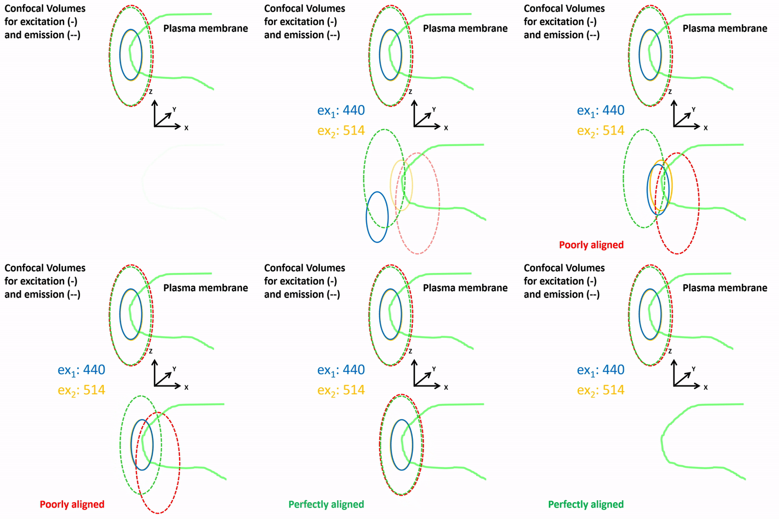
Perfect alignment is a key prerequisite for biomedical imaging in general, and quantitative molecular microscopy with pixel based analysis in particular. Since subsequent data correction is very limited we put substancial effort into the allignment and calibration of our setups. For that we developed calibration routines and procedures which we run on a regular basis.
Important Publications:
- Müller FE, Cherkas V, Stopper G, Caudal LC, Stopper L, Kirchhoff F, Henneberger V, Ponimaskin EG, Zeug A. Deciphering spatio-temporal fluorescence changes using multi-threshold event detection (MTED). bioRxiv 2020.12.06.413492.
- Butzlaff M, Weigel A, Ponimaskin E, Zeug A. eSIP: A Novel Solution-Based Sectioned Image Property Approach for Microscope Calibration. PLoS One. 2015 Aug 5;10(8):e0134980.
- Stawarski M, Rutkowska-Wlodarczyk I, Zeug A, Bijata M, Madej H, Kaczmarek L, Wlodarczyk J. Genetically encoded FRET-based biosensor for imaging MMP-9 activity. Biomaterials. 2014 Feb;35(5):1402-10.
- Zeug A, Woehler A, Neher E, Ponimaskin EG. Quantitative intensity-based FRET approaches--a comparative snapshot. Biophys J. 2012 Nov 7;103(9):1821-7.
- Weigel A, Schild D, Zeug A. Resolution in the ApoTome and the confocal laser scanning microscope: comparison. J Biomed Opt. 2009 Jan-Feb;14(1):014022.
- Neher RA, Mitkovski M, Kirchhoff F, Neher E, Theis FJ, Zeug A. Blind source separation techniques for the decomposition of multiply labeled fluorescence images. Biophys J. 2009 May 6;96(9):3791-800.
- Wlodarczyk J, Woehler A, Kobe F, Ponimaskin E, Zeug A, Neher E. Analysis of FRET signals in the presence of free donors and acceptors. Biophys J. 2008 Feb 1;94(3):986-1000.