New angiography system enables interventions even on small brain vessels

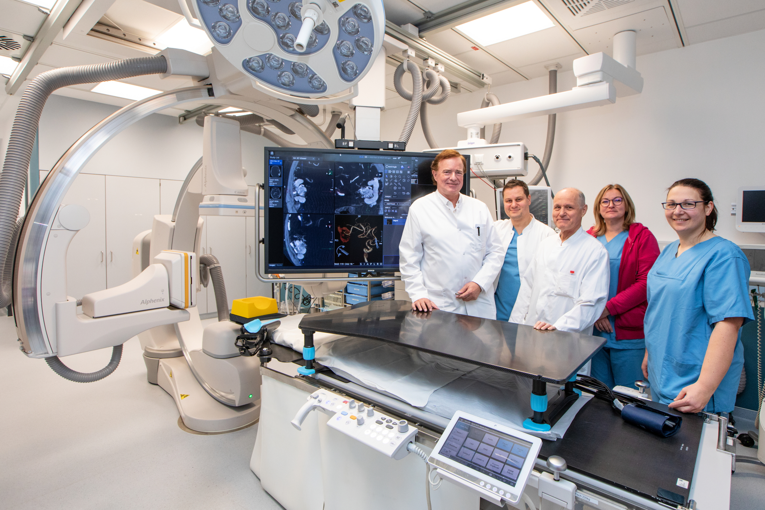
Professor Lanfermann (left) and his team are delighted about the new angiography equipment. Copyright: Karin Kaiser / MHH
Stroke, cerebral hemorrhage, aneurysm, vasoconstriction: For these diseases of the blood vessels, angiography imaging not only provides physicians with the best possible diagnoses. In addition, minimally invasive and often life-saving treatments can also be carried out during angiography. A new, extremely powerful angiography system from Canon was put into operation today at the Institute of Diagnostic and Interventional Neuroradiology at Hannover Medical School (MHH). The device has a very high-resolution image detector, which enables optical magnification of the brain vessels by a factor of two. Effective methods for reducing radiation exposure are also available. This means that patients can be offered even better treatment, for example in the event of a severe stroke.
Thrombectomy removes blood clots
Every year, the Interventional Neuroradiology department treats around 700 people with various vascular diseases of the brain, spine or spinal cord as well as tumors of the head and neck. Around 220 of these are patients who have suffered a stroke. The aim is to remove the blood clot in the cerebral vessel responsible for the stroke as quickly as possible in order to restore the blood supply to the brain. If this cannot be achieved with medication, a so-called thrombectomy can be performed using angiography imaging. "This involves inserting a telescopic catheter system from the groin or forearm into the affected cerebral vessel and either aspirating the clot or pulling it out," explains Dr. Friedrich Götz, Head of Interventional Neuroradiology.
Proven treatment becomes even more effective
"The fastest possible thrombectomy from vessels supplying the brain has significantly improved stroke treatment in recent years," says Professor Dr. Heinrich Lanfermann, Director of the Institute of Diagnostic and Interventional Neuroradiology. Until now, the procedure has mainly been used when large blood vessels in the brain are blocked by a clot. The new angiography system now provides excellent additional options in this area: Thanks to the high magnifications in excellent image quality, smaller vessels that supply the speech center in the brain, for example, can also be depicted very well, so that thrombectomies can also be carried out there under the best possible control. "This is a great benefit for the patients concerned," emphasizes Professor Lanfermann.
New possibilities, less X-ray radiation
Another technical innovation of the device is the so-called VASO-CT. It creates high-contrast, high-resolution cross-sectional images of even very small vessels.This is advantageous for the treatment of aneurysms, for example aneurysms, i.e. vascular protrusions: "The images allow us to see exactly whether a stent inserted for stabilization is fully in contact with the vessel wall or whether we still need to optimize the position," explains senior physician Dr. Omar Abu-Fares. The neuroradiologist is also enthusiastic about another innovation enthusiastic. "The device works with less X-ray radiation. It only illuminates the selected area with the necessary dose of radiation, while the surrounding tissue is less radiation and is therefore spared."
More flexibility in emergencies
The new angiography system fits perfectly into the existing technical technical equipment at the institute. It is located in the immediate tomography and the extended and newly equipped anaesthesia induction rooms. This means that patients do not have to be transported far during not have to be transported far during treatment. "The best thing, however, is that we now have not just one, but two angiography units with identical technical equipment," explains Dr. Götz. "With these two devices, we are now extremelyflexible. We can carry out planned interventions and also treattreat emergencies at the same time."
Close collaboration with other specialist areas
Diagnostic and interventional neuroradiology does not work on its own, but in closeon its own, but in close cooperation with other specialties such as neurology neurology, neurosurgery, anaesthesia, angiology, vascular surgery and ear, nose and throat medicine. For example, neuroradiology is a permanent partner of the supra-regionalcertified stroke unit, i.e. the stroke unit of the Clinic for Neurology. Neurology Clinic. It offers all diagnostic and therapeutic procedures for stroke patients around the clock, 365 days a year.and treatment procedures for stroke patients - including thrombectomy and including thrombectomy. "Especially in stroke treatment, the new angiographythe new angiography system will bring us another big step forward," says Professor Lanfermann is certain.
Text: Tina Götting