AG "Quantitatives und Metabolisches Neuroimaging"
Leitung: Prof. Dr. med. Dr. rer. nat. X. Ding
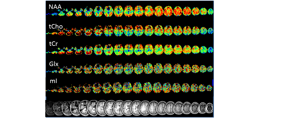
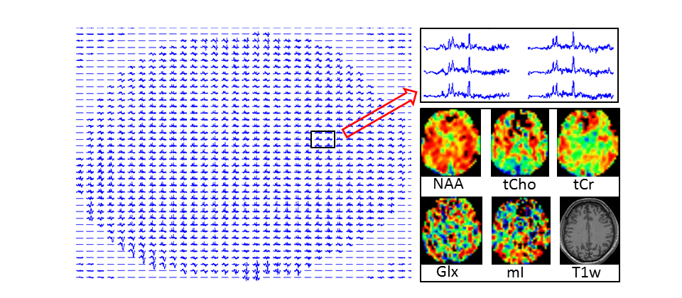
Die AG Ding beschäftigt sich mit der Etablierung neuer MR-Verfahren, um die mikrostrukturellen und metabolischen Veränderungen im Gehirn in vivo zu detektieren und zu erforschen. Ein Forschungsschwerpunkt der AG ist das quantitative MR-Imaging (qMRI). Es wurden verschiedene quantitative Mapping-Verfahren als Tools für die wissenschaftliche Forschung und klinische Anwendung entwickelt bzw. optimiert, wie z.B. T1-, T2-, T2*- und T2‘- Mapping. In Kombination mit DTI ist das qMRI häufig in der Lage, die subtilen und deshalb in der konventionellen MRT nicht sichtbaren pathologischen Veränderungen bei verschiedenen neurodegenerativen Erkrankungen festzustellen. Ein weiteres Forschungsgebiet ist die 1H-MR Spektroskopie (1H-MRS). Mit 1H-MRS kann man die Konzentrationen von Metaboliten - wie dem N-Acetyl-Aspartat (NAA) als Marker für Integrität von neuronalem Gewebe, dem gesamten Cholin (Cho) als Marker des Membran-phospholipid-metabolismus, dem gesamten Kreatin (tCr) als Marker für den Energiemetabolismus oder dem Myo-Inositol (mI) als Marker für die Gliosis und als Osmolyt sowie dem Glutamin/Glutamat (Glx) als Neurotransmitter - in vivo im Gehirn bestimmen und Informationen über Neurometabolismus gewinnen. Allerdings können die bisherigen 1H-MRS Verfahren jedes Mal nur ein kleines Hirnareal untersuchen, und zwar mit einer relativen langen Untersuchungszeit. Dadurch ist der Einsatz von 1H-MRS limitiert. Als neuesten Erfolg hat die AG mit Hilfe einer von der DFG finanziell unterstützten internationalen Kooperation ein weltweit neuartiges Verfahren zum Ganzhirn-Magnetresonanz-Spektroskopie-Imaging (wbMRSI) in der MHH erfolgreich aufgebaut. Mit diesem wbMRSI können sogenannte Metaboliten-Maps für das gesamte Gehirn erstellt werden (Abbildung 1), die detaillierte Information über Metaboliten-Verteilungen im Gehirn enthalten (Abbildung 2). Mit diesem wertvollen Werkzeug können wir ganz neue Aspekte in der Erforschung des Neurometabolismus einbringen und neue/tiefe Kenntnisse z.B. über die physiologischen Alterungsprozesse in Gesunden oder über die pathologischen neurodegenerativen Prozesse in Patienten gewinnen.


As a physicist and medical doctor she has been/is principal investigator of different research projects founded by German Research Foundation (DFG) and Federal Ministry of Education and Research (BMBF), for example, the project “Classifying Leukodystrophies with unknown defects according to neuroradiological criteria: applications of MRI patterns and MRS characteristics” (BMBF), or the project “Assessment of metabolic and microstructural correlates in aging human brain and in patients by using an innovative whole brain 1H magnetic resonance spectroscopic technique in combination with quantitative magnetic resonance imaging” (DFG). She is experienced in Mössbauer spectroscopy, MR imaging and MR spectroscopy, and combines both professions - physics and medicine - in the scientific research.
- Assessment of metabolic and microstructural correlates in patients with Parkinson’s disease and with major depression by use of whole brain 1H magnetic resonance spectroscopic technique in combination with quantitative magnetic resonance imaging
- Assessment of metabolic and microstructural correlates in patients with Post-COVID-Syndrome
- Quantitative MR brain morphological measurements in normal aging human and in patients
- Physiological and pathological microstructural alterations in aging human brain studied with diffusion tensor imaging
Recent publications:
1. Hennemann AK, Mahmoudi N, Döring K, Lanfermann H, Weissenborn K, Dirks M, Ding XQ. Evidence of Impaired Neuroimmune System in Post-COVID Syndrome-A Whole Brain Magnetic Resonance Spectroscopy Study. Journal of medical virology. 2025 Dec;97(12):e70762.
2. Mahmoudi N, Dadak M, Bronzlik P, Maudsley AA, Sheriff S, Lanfermann H, Ding XQ. Microstructural and Metabolic Changes in Normal Aging Human Brain Studied with Combined Whole-Brain MR Spectroscopic Imaging and Quantitative MR Imaging. Clin Neuroradiol. 2023 Dec;33(4):993-1005.
3. Klietz M, Mahmoudi N, Maudsley AA, Sheriff S, Bronzlik P, Almohammad M, Nösel P, Wegner F, Höglinger GU, Lanfermann H, Ding XQ. Whole-Brain Magnetic Resonance Spectroscopy Reveals Distinct Alterations in Neurometabolic Profile in Progressive Supranuclear Palsy. Movement disorders : official journal of the Movement Disorder Society. 2023 Aug;38(8):1503-14.
4. Reichardt JL, Dirks M, Wirries AK, Pflugrad H, Nösel P, Haag K, Lanfermann H, Wedemeyer H, Potthoff A, Weissenborn K, Ding XQ. Brain metabolic and microstructural alterations associated with hepatitis C virus infection, autoimmune hepatitis and primary biliary cholangitis. Liver international : official journal of the International Association for the Study of the Liver. 2022 Apr;42(4):842-52.
5. Stapel B, Nösel P, Heitland I, Mahmoudi N, Lanfermann H, Kahl KG, Ding XQ. In vivo magnetic resonance spectrometry imaging demonstrates comparable adaptation of brain energy metabolism to metabolic stress induced by 72 h of fasting in depressed patients and healthy volunteers. Journal of psychiatric research. 2021 Nov;143:422-8.
6. Klietz M, Elaman MH, Mahmoudi N, Nösel P, Ahlswede M, Wegner F, Höglinger GU, Lanfermann H, Ding XQ. Cerebral Microstructural Alterations in Patients With Early Parkinson's Disease Detected With Quantitative Magnetic Resonance Measurements. Frontiers in aging neuroscience. 2021;13:763331.
7. Fu T, Kobeleva X, Bronzlik P, Nösel P, Dadak M, Lanfermann H, Petri S, Ding XQ. Clinically Applicable Quantitative Magnetic Resonance Morphologic Measurements of Grey Matter Changes in the Human Brain. Brain sciences. 2021 Jan 5;11(1).
8. Ahlswede M, Nösel P, Maudsley AA, Sheriff S, Mahmoudi N, Bronzlik P, Lanfermann H, Ding XQ. Alterations of Striato-Thalamic Metabolism in Normal Aging Human Brain-An MR Metabolic Imaging Study. Metabolites. 2021 Jun 9;11(6).
9. Pflugrad H, Nösel P, Ding X, Schmitz B, Lanfermann H, Barg-Hock H, Klempnauer J, Schiffer M, Weissenborn K. Brain function and metabolism in patients with long-term tacrolimus therapy after kidney transplantation in comparison to patients after liver transplantation. PloS one. 2020;15(3):e0229759.
10. Kahl KG, Atalay S, Maudsley AA, Sheriff S, Cummings A, Frieling H, Schmitz B, Lanfermann H, Ding XQ. Altered neurometabolism in major depressive disorder: A whole brain (1)H-magnetic resonance spectroscopic imaging study at 3T. Progress in neuro-psychopharmacology & biological psychiatry. 2020 Jul 13;101:109916.
11. Fu T, Klietz M, Nösel P, Wegner F, Schrader C, Höglinger GU, Dadak M, Mahmoudi N, Lanfermann H, Ding XQ. Brain Morphological Alterations Are Detected in Early-Stage Parkinson's Disease with MRI Morphometry. Journal of neuroimaging : official journal of the American Society of Neuroimaging. 2020 Nov;30(6):786-92.
12. Dirks M, Pflugrad H, Tryc AB, Schrader AK, Ding X, Lanfermann H, Jäckel E, Schrem H, Beneke J, Barg-Hock H, Klempnauer J, Falk CS, Weissenborn K. Impact of immunosuppressive therapy on brain derived cytokines after liver transplantation. Transplant immunology. 2020 Feb;58:101248.
13. Schmitz B, Pflugrad H, Tryc AB, Lanfermann H, Jackel E, Schrem H, Beneke J, Barg-Hock H, Klempnauer J, Weissenborn K, Ding XQ. Brain metabolic alterations in patients with long-term calcineurin inhibitor therapy after liver transplantation. Alimentary pharmacology & therapeutics. 2019 Jun;49(11):1431-41.
14. Maghsudi H, Schutze M, Maudsley AA, Dadak M, Lanfermann H, Ding XQ. Age-related Brain Metabolic Changes up to Seventh Decade in Healthy Humans : Whole-brain Magnetic Resonance Spectroscopic Imaging Study. Clin Neuroradiol. 2019 Jul 26.
15. Maghsudi H, Schmitz B, Maudsley AA, Sheriff S, Bronzlik P, Schutze M, Lanfermann H, Ding X. Regional Metabolite Concentrations in Aging Human Brain: Comparison of Short-TE Whole Brain MR Spectroscopic Imaging and Single Voxel Spectroscopy at 3T. Clin Neuroradiol. 2019 Jan 18.
16. Klietz M, Bronzlik P, Nosel P, Wegner F, Dressler DW, Dadak M, Maudsley AA, Sheriff S, Lanfermann H, Ding XQ. Altered Neurometabolic Profile in Early Parkinson's Disease: A Study With Short Echo-Time Whole Brain MR Spectroscopic Imaging. Frontiers in neurology. 2019;10:777.
17. Goede LL, Pflugrad H, Schmitz B, Lanfermann H, Tryc AB, Barg-Hock H, Klempnauer J, Weissenborn K, Ding XQ. Quantitative magnetic resonance imaging indicates brain tissue alterations in patients after liver transplantation. PloS one. 2019;14(9):e0222934.
18. Dirks M, Pflugrad H, Tryc AB, Schrader AK, Ding X, Lanfermann H, Jackel E, Schrem H, Beneke J, Barg-Hock H, Klempnauer J, Falk CS, Weissenborn K. Impact of immunosuppressive therapy on brain derived cytokines after liver transplantation. Transplant immunology. 2019 Oct 24:101248.
19. Dirks M, Haag K, Pflugrad H, Tryc AB, Schuppner R, Wedemeyer H, Potthoff A, Tillmann HL, Sandorski K, Worthmann H, Ding X, Weissenborn K. Neuropsychiatric symptoms in hepatitis C patients resemble those of patients with autoimmune liver disease but are different from those in hepatitis B patients. Journal of viral hepatitis. 2019 Apr;26(4):422-31.
20. Schmitz B, Wang X, Barker PB, Pilatus U, Bronzlik P, Dadak M, Kahl KG, Lanfermann H, Ding XQ. Effects of Aging on the Human Brain: A Proton and Phosphorus MR Spectroscopy Study at 3T. Journal of neuroimaging : official journal of the American Society of Neuroimaging. 2018 Jul;28(4):416-21.
21. Pflugrad H, Schrader AK, Tryc AB, Ding X, Lanfermann H, Jackel E, Schrem H, Beneke J, Barg-Hock H, Klempnauer J, Weissenborn K. Longterm calcineurin inhibitor therapy and brain function in patients after liver transplantation. Liver transplantation : official publication of the American Association for the Study of Liver Diseases and the International Liver Transplantation Society. 2018 Jan;24(1):56-66.
22. Ding XQ, Maudsley AA, Schweiger U, Schmitz B, Lichtinghagen R, Bleich S, Lanfermann H, Kahl KG. Effects of a 72 hours fasting on brain metabolism in healthy women studied in vivo with magnetic resonance spectroscopic imaging. Journal of cerebral blood flow and metabolism : official journal of the International Society of Cerebral Blood Flow and Metabolism. 2018 Jan 01;38(3):469-78.
23. Kahl KG, Georgi K, Bleich S, Muschler M, Hillemacher T, Hilfiker-Kleinert D, Schweiger U, Ding X, Kotsiari A, Frieling H. Altered DNA methylation of glucose transporter 1 and glucose transporter 4 in patients with major depressive disorder. Journal of psychiatric research. 2016 May;76:66-73.
24. Eylers VV, Maudsley AA, Bronzlik P, Dellani PR, Lanfermann H, Ding XQ. Detection of Normal Aging Effects on Human Brain Metabolite Concentrations and Microstructure with Whole-Brain MR Spectroscopic Imaging and Quantitative MR Imaging. AJNR Am J Neuroradiol. 2016 Mar;37(3):447-54.
25. Ding XQ, Maudsley AA, Sabati M, Sheriff S, Schmitz B, Schutze M, Bronzlik P, Kahl KG, Lanfermann H. Physiological neuronal decline in healthy aging human brain - An in vivo study with MRI and short echo-time whole-brain (1)H MR spectroscopic imaging. NeuroImage. 2016 Aug 15;137:45-51.