
Interesting cases
A 26-year-old female patient with no previous illnesses had hallux valgus surgery in an orthopaedic Clinical Department.
During the first operation, the wound could not be closed and a soft tissue defect remained. For this reason, the patient underwent VAC therapy, which was followed by several more. As the wound could not be closed even after 10 weeks, the osteosynthesis material was removed. However, a soft tissue defect remained on the left foot, which is why VAC therapy was initiated again.
The patient was then presented to the Clinical Department of Plastic, Aesthetic, Hand and Reconstructive Surgery.
You can read about the further procedure in the following article:
Closure of a large tissue defect after hallux valgus surgery - Best Wound Practice
A 60-year-old man was found unconscious in a house fire and admitted to the Center for Severe Burns with more than 40% of his body surface burned. On admission, the patient was already suffering from circulatory instability and severe pulmonary damage due to inhalation trauma. Global lung failure became apparent early on, which required lung replacement therapy. Thanks to interdisciplinary collaboration with the cardiothoracic surgery department, extracorporeal membrane oxygenation (ECMO) could be applied in good time and the lungs relieved. The 2b and 3rd degree burns required surgical treatment, which achieved complete defect coverage after 9 operations. The lung replacement procedure was transferred to controlled ventilation therapy and weaning from the respirator. Early rehabilitation in the intensive care unit led to full mobilization after a total of 85 days.
Despite the prognostically unfavourable burn severity, the organ failure of the lungs and the age (predictive outcome: 90% mortality risk), the poor prognosis was counteracted by targeted plastic surgery burn medicine and interdisciplinary collaboration with cardiothoracic surgery (ECMO), physical medicine and anaesthesiology, and the patient was discharged after 12 weeks to specialized rehabilitation for burn patients in Bad Klosterlausnitz.
Successful treatment of facial nerve palsy using microsurgical crossface nerve transplantation
Back in 2015, a 14-year-old female patient presented with unilateral, peripheral, complete facial nerve palsy. At the age of 4, she had suffered a domestic accident with a retroauricular hematoma and subsequent hearing loss and paralysis of the right facial nerve. Relief of the hematoma by ENT colleagues in the acute stage did not improve the findings. Following a detailed medical history and clinical findings, we initially indicated exploration and neurolysis of the facial nerve on the damaged side, as spontaneous motor activity was still present on the affected side of the face. This was carried out without complications, but did not lead to any improvement in the findings.
Together with the patient and her parents, we discussed all the options for improving facial expressions. In addition to static and dynamic mimic replacement operations, the main focus was on facial reanimation using a cross-face nerve graft, as the preserved but uncontrollable spontaneous motor function promised the prospect of reinnervation. A nerve graft (sural nerve) can be used to create a connection between the intact part of the healthy side and the damaged contralateral facial nerve using a microsurgical technique from the healthy side using the so-called crossover technique. With a latency for the formation of the axons (approx. 1 mm per day) to the opposite side, reinnervation of the paralyzed half of the face can be achieved. Thanks to our Clinical Department's expertise in peripheral nerve surgery, we were able to offer the young girl the most effective and scar-saving reconstruction possible. The nerve transplant was successfully performed in July 2016. At the clinical follow-up in December, there were already clear signs of reinnervation of the right side of the face. Through ergo- and physiotherapeutic stimulation and exercise treatment, a visible success was achieved for the patient, which represents a major gain in quality of life.
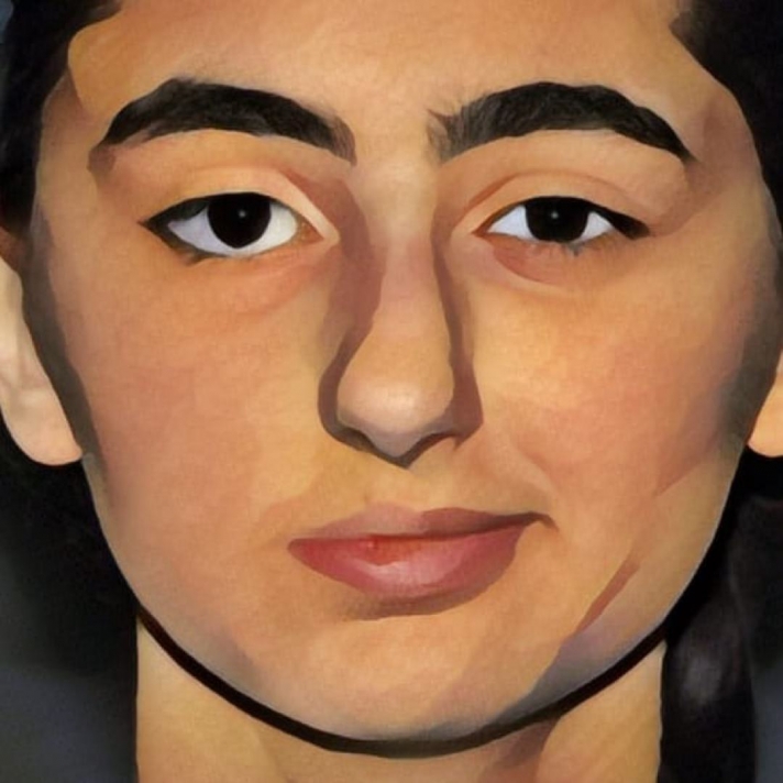
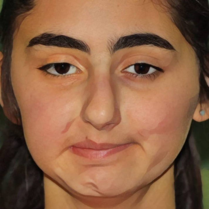
Schematized representation of the photo documentation with complete paresis of the right half of the face (A) in July 2016 and 5 months after cross-face transplantation with incipient mimic activation of the right corner of the mouth and the right lower eyelid (B).
In our special consultation hour for paralysis and loss of function of visible functions of the face and extremities, we are happy to advise your patients on the possibilities of innovative, microsurgical methods or functional replacement operations.
In June, an 85-year-old female patient presented to our emergency department with a garlic burn. According to the patient, the reason for using this home remedy was joint pain in her left knee and back pain.
The use of garlic extract for the treatment of multiple complaints and diseases in internal medicine and rheumatology is traditional and has been used for over 5000 years. Garlic is used in various forms in both European and traditional Chinese medicine (TCM).
In our case, the patient suffered deep dermal burns on 6 % of her body surface, so that the wounds had to be surgically debrided with subsequent split-thickness skin transplantation. The patient used uncooked garlic extract, which can have a corrosive effect on prolonged skin contact and can cause extensive damage to the skin. The patient has since been discharged with her skin grafts well healed and mobilized.
Only 17 sources worldwide can be found as case reports and statements in the literature, which is why this "rarity" calls for further investigations. We will continue to investigate the effect of garlic extract on the skin under scientific conditions, in different doses and processing, and will report the results in one of the next newsletters.
In February 2016, an 18-year-old forestry worker suffered a complete dislocation of his legal arm in an accident at work. The arm got caught in a cable winch and was completely amputated due to the enormous forces, in the sense of a laceration-crushing injury.
The arm was successfully replanted in a 9-hour operation.
In further operations, the lost skin and soft tissue mantle was restored by microsurgical tissue transfer from the right thigh to the right upper arm.
A free Mutton-Chop flap was used for the reconstruction. The initial description goes back to a publication from our Clinical Department. This is based on the neurovascular supply of the lateral circumflex femoral artery and the femoral innervation of the quadriceps femoris. Including the rectus femoris and tensor fascia latae muscles and an extended skin island (in the sense of an anterior-lateral high flap/ALT) of up to 40 x 20 cm, a myocutaneous, neurovascular flap plasty can thus adequately cover large defects and form the basis of a functional reconstruction.
Further operations for functional reconstruction are planned.