Preformation/bone replacement

Defects in the bony structures of the facial skull, such as the partial or complete loss of bone sections, are a common problem in maxillofacial surgery. These can occur due to congenital defects, as a result of accidents or during the removal of tumors. In addition to aesthetic problems, the consequences of these defects include functional limitations, e.g. when chewing, because muscle attachments are missing or part of the lower jaw has had to be removed. In order to close these bone defects, various bone replacement materials that are similar in structure and composition to natural bone are used for smaller defects.
As part of our research, we are looking for new replacement materials and trying to better characterize already known replacement materials. We are also trying to improve the ingrowth of the body's own tissue and blood vessels into these materials.
In the case of larger defects, the replacement materials can no longer be used - the patient's own tissue must be used. For this purpose, bone (sometimes with soft tissue structures) is removed from one part of the patient's body and used. This procedure creates an additional defect site in the body, which can lead to further health problems.
Tissue engineering (TE)
In order to avoid the above-mentioned problems, so-called "tissue engineering" is used in preoperative planning, whereby implants tailored to the patient and the specific defect situation are developed. These constructs are grown from the patient's cells in the laboratory and do not cause a secondary removal defect.
A serious problem with defect treatment using TE constructs is the limited survival rate of the constructs after implantation, as there is no connection to the microcirculation in the defect area. The basis of oxygen and nutrient supply is not sufficient to supply constructs of clinically relevant size.
We are trying to develop strategies to improve the survival of the constructs and are investigating the development of the vascular system. Various models are available with which changes in the vascular system can be directly tracked and quantified.
" back to the research page
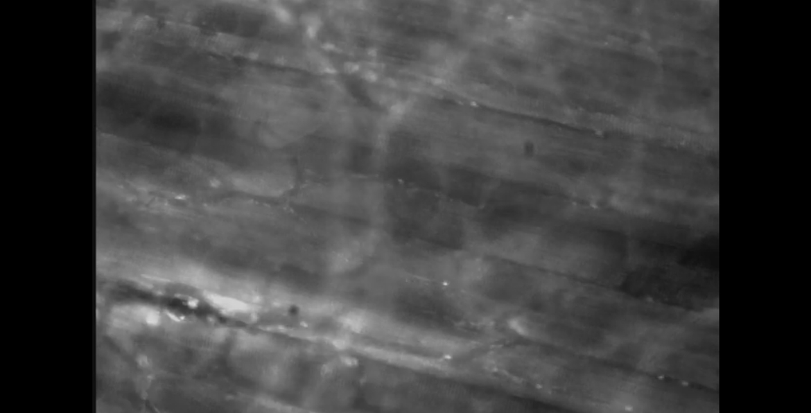
Intravital fluorescence microscopy of the microcirculation in a "tissue engineering" construct