Researchers from Hanover, Mainz, Wuppertal and Witten develop high-resolution tomography - application conceivable for Covid-19, Alzheimer's and cancer.

Professor Dr Danny Jonigk. Copyright: Karin Kaiser/MHH
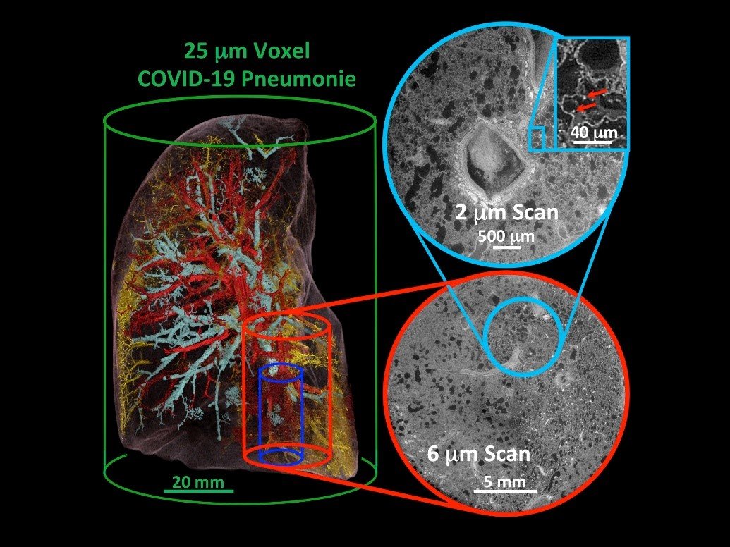
Three-dimensional segmentation and reconstruction of a lung with Covid-19 pneumonia. Individual thrombi (yellow), vascular changes (red) and airway changes (turquoise) can be visualised via the high-resolution method of Hierarchical Phase-Contrast Tomography (HiP-CT).
Status: 17 February 2021
Researchers from Hanover Medical School (MHH), Mainz University Hospital and HELIOS University Hospital Wuppertal at Witten/Herdecke University are leading an international, multidisciplinary consortium that makes high-resolution, three-dimensional X-ray images of the human body possible. With a maximum resolution of up to 300 nanometres, they are equivalent to one tenth of the diameter of a human hair. "The networking between radiology, pathology and molecular diagnostic approaches will take on enormous clinical significance in the future," emphasises pathologist Prof. Dr. Danny Jonigk, MHH Institute of Pathology. "With the high-resolution three-dimensional imaging, we will be able to better understand tissue damage in the lungs in covid-19," says PD Dr Maximilian Ackermann, Institute of Pathology at Helios University Hospital Wuppertal, University of Witten/Herdecke, and at the Institute of Functional and Clinical Anatomy at the University Medical Center Mainz.
Both scientists are certain that this novel, high-resolution imaging technique will also provide fascinating new insights into other diseases, such as cancer or Alzheimer's dementia. They are currently working under high pressure with their colleagues in England and France to use this innovative technique to spatially characterise and understand the tissue damage of covid 19 pneumonia. The Chan Zuckerberg Initiative (CZI) of Facebook founder Mark Zuckerberg and his wife, the physician Dr. Priscilla Chan, is funding this project with one million US dollars.
Making the clinically known CT a hundred times more accurate
Three-dimensional imaging is familiar from conventional clinically used computed tomography (CT). However, high-resolution micro-computed tomography using synchrotron radiation provides much higher resolutions. This technology is based on high-energy X-rays generated by synchrotron particle accelerators. The international team of this worldwide unique project entitled "Human Organ Project" is composed of physicians from the European Synchrotron Facility ESRF in Grenoble and bioinformatics as well as biomechanics experts from University College London (UCL) in addition to the physicians from Germany. For the first time, the scientists can make use of a recently reconstructed radiation source at the ESRF in Grenoble. This "Extremely Brilliant Source" (EBS) is the world's first high-energy synchrotron source of the fourth generation and currently the brightest X-ray source in the world. The project promises images of the entire human body with a resolution of two micrometres, over a hundred times better than a CT scanner. "We were very pleased that the Chan Zuckerberg Initiative is funding our translational approach," explains PD Dr Maximilian Ackermann.
The Chan Zuckerberg Initiative (CZI) was launched in December 2015 by Mark Zuckerberg, founder of Facebook, and his wife, the paediatrician Dr Priscilla Chan, in East Palo Alto. The Chan Zuckerberg Initiative promotes engineering, basic biomedical research and artificial intelligence.