Anatomy
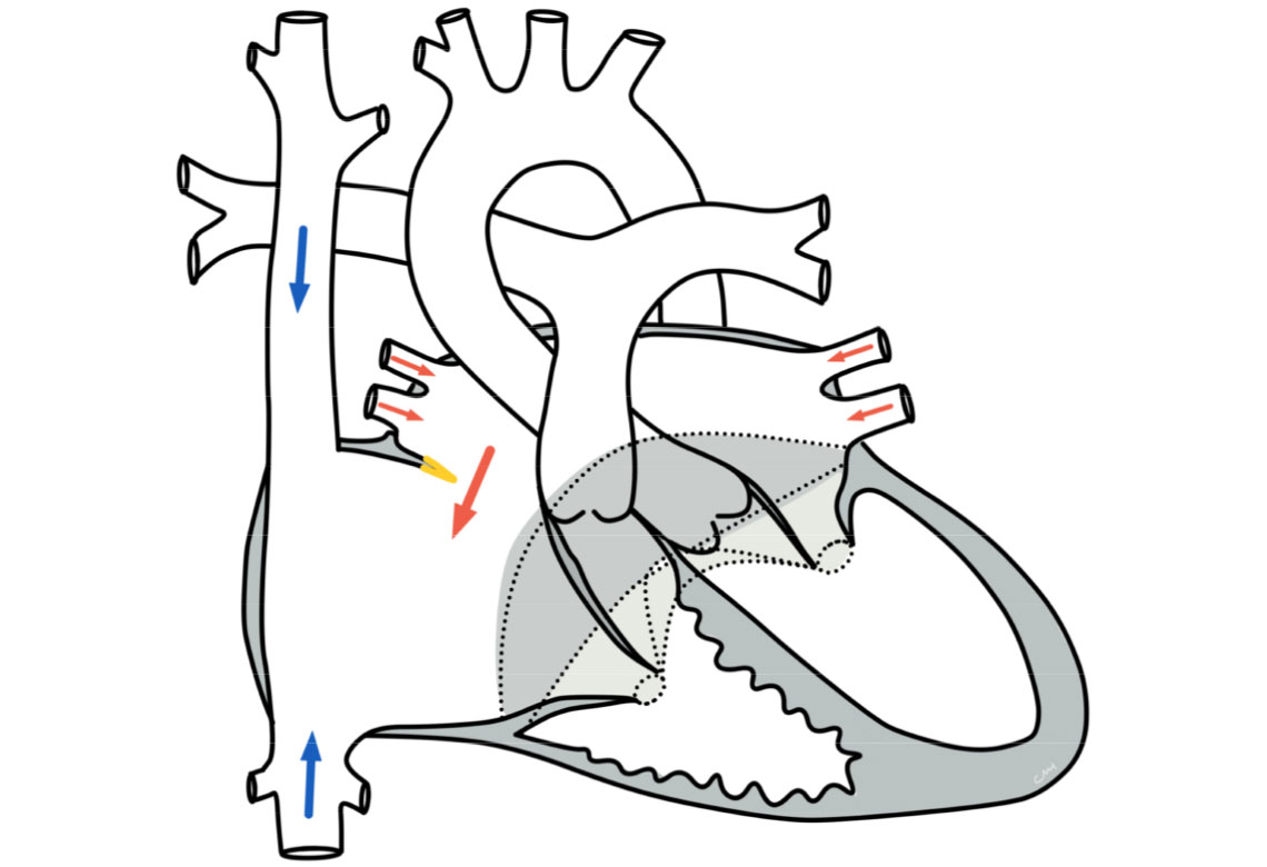
Anatomically, the two ventricles (atria) are separated from each other by a septum. In an ASD I, there is a connection between the left and right atrium through an interruption in this septum.
The defect is located far down in the atrial septum near the atrioventricular valves (AV valves). The size of the defects varies. The left and right AV valves have a common valve ring and this defect is often associated with a cleft in the left-sided AV valve, which can lead to leakage of this valve.
Physiology and hemodynamics
Depending on the size of the connection, more or less blood flows through the defect. As there is a slightly higher pressure in the left atrium, blood flows through the defect from the left into the right atrium (left-to-right shunt), from there into the right ventricle and then into the pulmonary vessels. This can lead to a strain on the right heart and to an over-perfusion of the lungs.
Unlike a ventricular septal defect, however, an atrial septal defect usually does not pose a risk of pulmonary hypertension, as the pressure in the two atria is significantly lower than in the main chambers. Therefore, although an atrial septal defect increases the blood flow to the lungs, there is no significant pressure load.
Therapy for a primum-type atrial septal defect
Conservative
The need for treatment depends on the size of the defect and therefore on the effects on the circulation (hemodynamic relevance). An ASD I does not close by itself. In the case of small defects, the hemodynamic effects are not noticeable for a long time. However, there are also defects that are so large that the children are noticeably impaired at a very early age - sometimes even in infancy: the increased volume of blood flowing through the lungs puts a strain on the children and they become cardiac insufficiency. Rapid breathing, a rapid pulse, sweating under stress (such as when drinking or crying) and slow weight gain are noticeable. In these cases, drug therapy is started before surgical correction.
Surgical therapy
The definitive treatment is surgical correction. Interventional closure is not possible with this type of defect due to the position of the defect in relation to the AV valves. Careful planning with the aid of preliminary echocardiographic examinations is necessary to determine the size, location and haemodynamic relevance of the defect. The surgical closure is performed using the heart-lung machine, with an anterior approach through the chest. The defect is closed with a patch made of pericardial material or other tissue (synthetic or biomaterial). After the operation, the patient is treated for a short time in the intensive care unit before returning to the cardiology follow-up ward.
Prognosis for a primum-type atrial septal defect
Further course
After timely therapeutic closure, a normal physical capacity and an unimpaired lifestyle can be expected.