Range of courses

Copyright: Dr. N. Schaumann, Pathology, MHH
Range of courses


Admission:

The fully passed preliminary dental examination is a prerequisite for participation in all courses of the clinical study section, including the General and Special Pathology modules.


Learning objectives:

The teaching of pathology comprises the teaching of the most important characteristics of macroscopically and microscopically detectable pathological changes in cells, tissues and organs together with the underlying pathophysiological principles.


Topics of general pathology:

  1. Introduction to pathology
  2. Cell damage and adaptation reaction
  3. Circulatory disorders
  4. Acute inflammation
  5. Chronic inflammation
  6. Pathological immune reactions and inflammation in the head and neck area
  7. Tumor pathology I (general tumor pathology)
  8. Tumor pathology II (classification, metastasis)
  9. Tumor pathology III (molecular basics)
  10. Tumor pathology IV (tumors in the head and neck area)

Macroscopic specimens are demonstrated at the institute.


Topics of special pathology:

  1. Arteriosclerosis
  2. Acute myocardial infarction
  3. Cardiac callus after infarction
  4. Chronic pulmonary edema
  5. Pulmonary emphysema
  6. Lobar pneumonia - red hepatization
  7. Lobar pneumonia - grey hepatization
  8. Organizing pneumonia
  9. Interstitial pulmonary fibrosis
  10. Tuberculosis
  11. Liver fibrosis in chronic hepatitis
  12. Liver cirrhosis
  13. Glomerulonephritis
  14. Diabetic glomerulopathy
  15. Gastric ulcer
  16. Acute appendicitis
  17. Hodgkin's lymphoma
  18. Sarcoidosis
  19. Graves' disease
  20. Seborrheic keratosis
  21. Nevus cell nevus
  22. Basal cell carcinoma
  23. Malignant melanoma
  24. Mucoepidermoid carcinoma
  25. Adenoid cystic carcinoma
  26. Warthin tumor
  27. Fibroepithelial hyperplasia
  28. Squamous cell papilloma
  29. Apical granuloma
  30. Radicular cyst
  31. Follicular cyst
  32. Keratocyst
  33. Fibrous dysplasia
  34. Osteomyelitis of the jaw
  35. Giant cell pulis
  36. Odontoameloblastoma
  37. Ameloblastic fibroma
  38. Ameloblastoma
  39. Non-Hodgkin's lymphoma
  40. Kaposi's sarcoma
  41. Leukoplakia
  42. Squamous cell carcinoma, well differentiated
  43. Squamous cell carcinoma, poorly differentiated


Microscopic preparations are demonstrated during the microscopy course and microscoped independently by the students.


Course achievements:

Compulsory participation in the microscopy course, lectures and the institute observation.


Examination form:

Structured oral exam (SOE). Knowledge from the lecture, textbooks and pathology course is tested on two histological preparations using selected examples from the microscopy course and the lectures.


Courses offered in accordance with the new Dental Licensing Regulations


Admission:

The fully passed preliminary dental examination is a prerequisite for participation in all courses of the clinical study section, including the pathology module.


Learning objectives:

The teaching of pathology covers the most important characteristics of macroscopically and microscopically detectable pathological changes in cells, tissues and organs together with the underlying pathophysiological principles.


Topics of general pathology:

  1. Introduction to pathology
  2. Cell damage and adaptation reaction
  3. Circulatory disorders
  4. Acute inflammation
  5. Chronic inflammation
  6. Pathological immune reactions
  7. General tumor pathology I
  8. General tumor pathology II
  9. Benign diseases of the head and neck
  10. Malignant diseases of the head and neck

The topics are based on the IMPP subject catalog dentistry (GK ZM) from 2023 for the new dental licensing regulations.

Theoretical content of the above-mentioned subject areas is taught in the lecture. On three lecture dates, the events take place in the microscopy room, which also gives students practical experience in microscopy of relevant clinical pictures.


Coursework:

Compulsory participation in the lecture
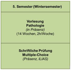
 

Examination form:

Written examination. Single-choice (from 5 possible answers), 30 questions, some with histological images. Online via the ILIAS platform. Knowledge from the lecture and textbooks will be tested.