MHH researchers are developing a novel membrane architecture with improved gas exchange for more compact ECMO systems and an artificial lung implant.
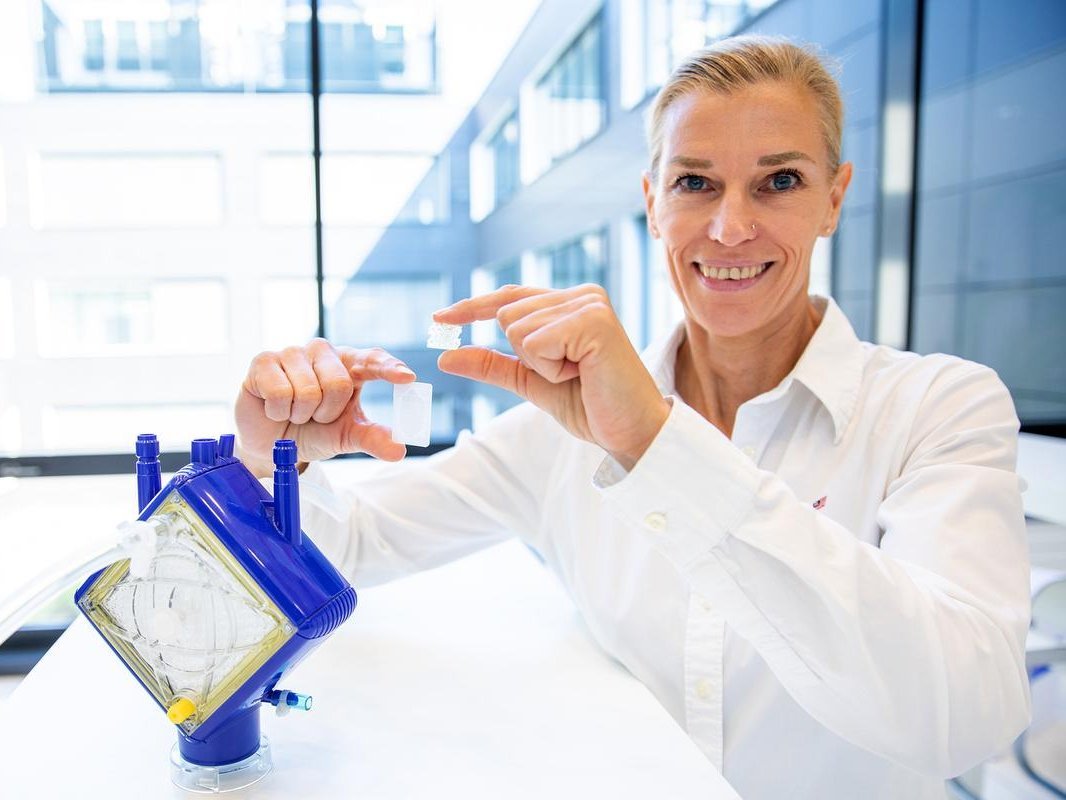
Smaller and more efficient: Prof. Dr. Bettina Wiegmann demonstrates the new TPMS membrane architecture (right) in comparison to the conventional hollow-fiber membrane bundles from an ECMO lung support system. Copyright: Karin Kaiser/MHH
In cases of severe lung disease, a lung transplant is often the last resort. However, donor organs are scarce. Until then, so-called ECMO ventilation systems can support lung function. Their architecture, consisting of hollow-fiber membranes, however, limits gas exchange, leads to uneven blood flow, and—together with the artificial surfaces—promotes blood clotting. A research team at MHH, in collaboration with RWTH Aachen University, has now developed a completely new, 3D-printable membrane architecture that improves not only oxygen delivery but also blood compatibility.
For people with severe lung diseases, a transplant of a healthy organ is often the only chance of survival. But donor lungs are in short supply. A new artificial lung could provide a solution. A team led by Prof. Dr. Bettina Wiegmann, an emergency medicine physician and specialist in cardiac surgery at the Clinical Department of Cardiac, Thoracic, Transplantation, and Vascular Surgery at Hannover Medical School (MHH), has been researching this since 2017 as part of the priority program “Towards an Implantable Lung” (“Towards an Implantable Lung”) of the German Research Foundation (DFG). Together with her research group at the Lower Saxony Centre for Biomedical Engineering, Implant Research and Development (NIFE), she is developing what is known as the biohybrid lung. This is based on extracorporeal membrane oxygenation (ECMO). In this lung support system, which is already in clinical use, blood is passed through plastic hollow-fiber membranes (HFM) that facilitate gas exchange.
Now, the scientist has taken a decisive step forward. Together with researchers from RWTH Aachen University, she has developed a novel membrane architecture that makes gas exchange much more efficient. This architecture can be produced easily and with precise fit using 3D printing, achieves significantly higher oxygen transfer, and at the same time enables a space-saving design. The method is not only of interest for the development of implantable biohybrid lungs. In the future, it could also make conventional ECMO systems more powerful and compact. The study has been published in the journal “Advanced Materials,” an internationally renowned journal for materials science.
New membrane architecture
The HFMs used in ECMO resemble tiny straws arranged side by side in parallel. However, this arrangement creates turbulence. As a result, the flow distribution is not ideal and limits the efficiency of gas exchange. In addition, there is an increased tendency for thrombosis to form at the interfaces between the blood and the artificial surfaces. “So far, we’ve only been able to use ECMO to bridge lung function for a certain period of time because the blood forms clots upon contact with the artificial surfaces,” explains Professor Wiegmann. She is therefore pursuing a different solution: a completely new membrane architecture based on so-called triply periodic minimal surfaces (TPMS). Unlike the densely packed HFM, the TPMS structure forms a continuous three-dimensional network. This allows the blood to be distributed more evenly, enabling more efficient gas exchange while also laying the foundation for more compact artificial lungs.
Up to 88 percent more oxygen
Similar to natural alveoli, TPMS structures have a very large surface area combined with a small volume. This ensures that as much surface area as possible is available for gas exchange within a very small space. In the human lung, approximately 100 to 140 square meters of respiratory surface area is compactly packed into 300 million alveoli. While the TPMS structures do not yet achieve this packing density, they avoid areas with low blood flow and reduce flow resistance. In addition, they can be easily and custom-manufactured using modern 3D printing. “We have optimized the TPMS architectures. As a result, we achieve up to 88 percent higher oxygen transfer than with conventional hollow-fiber membranes,” says Professor Wiegmann. “This means that in the future, the same or even better oxygen supply could be possible with significantly smaller artificial lungs.”
Goal: CT lung image as a printing template
Not only the architecture but also the material used opens up new possibilities. The TPMS membranes are made of a special silicone polymer. It is biocompatible, non-toxic, and chemically very stable. At the same time, the material is highly permeable to oxygen and carbon dioxide—a crucial prerequisite for efficient gas exchange. It can also be colonized by endothelial cells. These cells line our natural blood vessels and can regulate blood clotting. This could significantly improve the blood compatibility of future artificial lungs.
The long-term goal of this research is to use computed tomography (CT) scans of the damaged lung to create a “template” and use it to 3D-print custom-fit artificial lung segments or entire lungs. Seeded with the patient’s own or genetically modified endothelial cells, the artificial lungs could then be implanted in patients and permanently take over lung function. “The new TPMS structure has great potential even without endothelial cells,” the scientist emphasizes. “Even as a replacement for today’s hollow-fiber membranes, it could make ECMO systems more efficient, more compact, and better tolerated by the blood. In the long term, it also forms the basis for implantable biohybrid lungs.”
Expertise recognized in the U.S.
Professor Wiegmann now hopes that her work will continue to receive funding so that the implantable biohybrid lung will one day find its way into the Clinical Department—initially as a temporary solution until a lung transplant can be performed, and in the long term as a full-fledged organ replacement. A recent award from the U.S. also demonstrates that her research and expertise are widely recognized internationally. The American Society for Artificial Internal Organs (ASAIO) has inducted the scientist into the “Inaugural Fellow Class of 2026” in recognition of her research achievements, her international engagement, and her contributions to the further development of biohybrid organ support systems and extracorporeal therapeutic procedures. With this newly established and internationally renowned award, the professional society honors world-leading scientists for their outstanding contributions to the development of artificial organs, modern organ support systems, and innovative translational therapy concepts.
Text: Kirsten Pötzke
SERVICE:
The original paper, “From Fiber Bundles to Architected Membranes: Triply Periodic Minimal Surface Architectures for Biohybrid Artificial Lungs,” can be found here.
Further information can be found here.