A preclinical study by the MHH shows improved heart function following the transplantation of cardiac muscle cell aggregates.
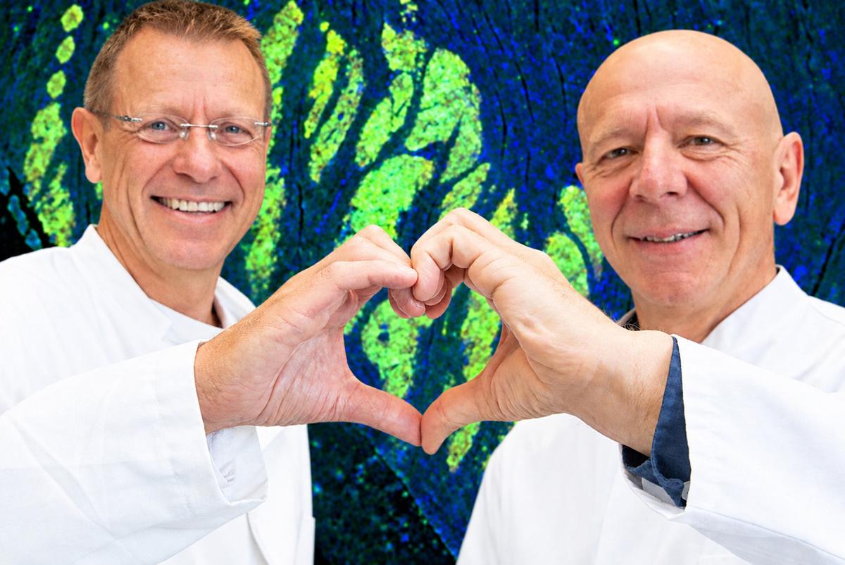
Concentrated research expertise in heart repair using laboratory-grown heart muscle cells: Prof. Dr. Ulrich Martin (left) and Prof. Dr. Robert Zweigerdt. Copyright: LEBAO; Karin Kaiser/MHH
In the case of a heart attack, the affected part of the heart muscle can no longer be supplied with oxygen and nutrients. As a result, the heart muscle cells (cardiomyocytes) die. Scar tissue forms, and heart function is impaired. This often leads to chronic heart failure—also known in medicine as cardiac insufficiency. It is one of the leading causes of death in the Western world. Prof. Dr. Ulrich Martin, director of the Leibniz Research Laboratories for Biotechnology and Artificial Organs (LEBAO) at Hannover Medical School (MHH), and Prof. Dr. Robert Zweigerdt, a cell biologist at LEBAO, aim to repair the damaged areas of the heart using biotechnologically produced heart muscle cells. What sets their approach apart is that they do not rely on individual cells, but rather on small clusters of human cardiomyocytes, which are produced in large quantities and injected directly into the heart muscle. They have now investigated this therapeutic concept in collaboration with and at the German Primate Center in Göttingen using a heart attack model in Javanese macaques. The preclinical study showed that the treatment clearly improved heart function after three months. The results have been published in the journal “Nature Communications.”
Cell aggregates form extensive muscle fibers in the heart
The researchers and their team used so-called human induced pluripotent stem cells (hiPSCs). These can be produced in the laboratory from “reprogrammed” somatic cells, such as blood cells, and can then give rise to any cell type in the human body—including heart muscle cells. Even during production in the laboratory in special bioreactors, these biotechnologically derived heart muscle cells grow into spherical cell aggregates. The hypothesis is that, compared to single cells, more of these aggregates remain in the heart and integrate better into the heart muscle.
In the current study, the researchers injected hiPSC-derived cardiac muscle cell aggregates (hiCMAs) into the hearts of primates that had previously undergone a heart attack. Three months after the procedure, extensive new human cardiac muscle fibers had formed. Although the cell transplant led to temporary cardiac arrhythmias, it had positive effects on heart function over time.
hiPSCs Have Their Own Heart Rate
There is a reason why the experiments were conducted on monkeys rather than, for example, mice. While mice and rats are generally suitable for many other animal studies, preclinical testing of this cell therapy concept cannot be performed in small-animal models. This is because their hearts are much smaller and have a significantly higher heart rate compared to humans. “This would not be compatible with human heart muscle cells, which have their own human heart rate, meaning that observations made here do not allow for conclusions to be drawn about humans,” explains Professor Zweigerdt. In large-animal models such as pigs, the heart is much more similar to that of humans in terms of size and metabolic processes, including heart rate. However, the animal’s immune system immediately recognizes the human cells as foreign and rejects them. In monkey hearts, on the other hand, the human donor cells can grow and survive largely without problems with appropriate immunosuppressive medication.
In their studies, the researchers tested hiCMAs derived from two different iPSC lines. “We found that only cardiomyocytes from one of the two lines had a positive effect on heart function, even though both lines resulted in human grafts of similar size in the monkey heart,” notes Professor Zweigerdt. “Our study suggests that the contraction of heart muscle cells in cell culture is linked to their potential to improve heart function. It is therefore important to examine the cell aggregates for their functions—including contraction force—prior to transplantation.”
Long-Term Therapies Using the Body’s Own Cells
There are already several ongoing clinical trials worldwide investigating therapies using cultured heart muscle cells. However, the stem cells used in these trials were not derived from the patient’s own cells but from donor cells. The problem: Similar to a donor organ, the immune response following cell transplantation must be suppressed with medication for the rest of the patient’s life. This poses an additional risk and limits the number of potential patients who could benefit from cell therapy using cardiac muscle cells. The MHH scientists are therefore pursuing a different long-term approach. They aim to transplant hiCMAs derived from the patients’ own, so-called autologous, blood cells. “A major advantage of this is that it does not require immunosuppression, which is associated with side effects,” notes Professor Martin. This could lead to greater acceptance, particularly among younger patients, and accelerate the development of the therapy despite higher costs.
Research on Stem Cell Therapy
For more than 20 years, Professor Martin and Professor Zweigerdt have been working with their team to develop cell therapies for the treatment of severe heart diseases. And they are making steady progress. As part of the iCARE project, the researchers have already successfully demonstrated in animal models that the cell aggregates actually integrate into heart tissue and improve organ function. In the EU-funded HEAL research project, they aim to refine the strategies for producing and administering the hiCM aggregates so that they can be used for cardiac therapy in humans. “Living cells are an extremely complex drug,” emphasizes Professor Martin. “While we cannot completely heal a pre-damaged heart with them, we can give it a sort of ‘fresh cell treatment’ that could improve its function in the long term.” However, before this therapy becomes part of standard care, it will still need to be tested in clinical trials for safety and tolerability. Yet the researchers are optimistic. In their new EU-funded project, iNDUCARE, they are already working on the prerequisites to make the complex production of the necessary autologous iPSCs simpler, faster, and more cost-effective.
The original publication, “Human iPSC-cardiomyocyte-aggregate cell therapy in non-human primates and correlation of heart recovery with contractile and electrophysiological cardiomyocyte properties,” can be found here.
Further information on biomedical research involving primates can be found here.
Text: Kirsten Pötzke