Anatomy
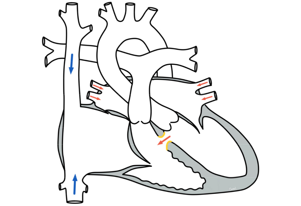
Anatomically, the two ventricles (atria) are separated from each other by a septum. In an ASD II, there is a connection between the left and right atrium through an interruption in this septum.
The defect is often located approximately in the middle of the atrial septum, in the area of the so-called foramen ovale (the opening between the two atria in the unborn child, which usually closes after birth). The size of the defects varies.
Physiology and hemodynamics
Depending on the size of the connection, more or less blood flows through the defect. As there is a slightly higher pressure in the left atrium, blood flows through the defect from the left into the right atrium (left-to-right shunt), from there into the right ventricle and then into the pulmonary vessels. This can lead to a strain on the right heart and to an over-perfusion of the lungs.
Unlike a ventricular septal defect, however, an atrial septal defect usually does not pose a risk of pulmonary hypertension, as the pressure in the two atria is significantly lower than in the main chambers. Therefore, although an atrial septal defect increases the blood flow to the lungs, there is no significant pressure load.
Therapy for a ventricular septal defect
Conservative
The therapy depends on the location and size of the defect and thus on the effects on the circulation (hemodynamic relevance). In many cases, no special therapy is necessary, as spontaneous closure of a ventricular septal defect occurs in approx. 30-40% of cases. These are usually small muscular defects that close themselves in the first year of life. Larger defects or defects that are still open after the first year of life very rarely close, but can become smaller. There are also defects with a special location (inlet defects and infundibular defects) that never close spontaneously, nor do they become smaller over time. If there are effects on the circulation, it is necessary to close the defect. Occasionally, drug therapy is used as a bridging measure (heart failure therapy) to allow the baby to grow (thrive).
Surgery and intervention
Depending on the location and size of the defect, the definitive therapy consists of surgery or intervention using catheter technology. Careful planning with the help of preliminary echocardiographic examinations is necessary to decide on the appropriate treatment procedure in order to visualize the size, position and shape of the defect. The surgical closure is performed using a heart-lung machine. The chest is opened in the middle. The position of the defect determines the access route in the heart. The defect is closed with a patch made of pericardial material or synthetic tissue. After the operation, the patient is treated in the intensive care unit for a short time before returning to the cardiology follow-up ward.
In the case of selected defects, closure can be performed in the catheter laboratory. This involves using a cardiac catheter to position a small umbrella or coil in the defect through a vascular access (sheath) inserted into the inguinal vessels so that the defect is closed. A drug is used to inhibit the formation of blood clots until the umbrella or coil has completely grown into the defect.
Prognosis for a ventricular septal defect
Further course
After spontaneous closure of the VSD, the children are heart-healthy and do not require any further check-ups. After timely therapeutic closure, normal physical resilience and an unimpaired lifestyle can be expected.