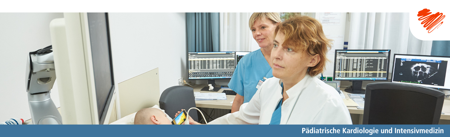
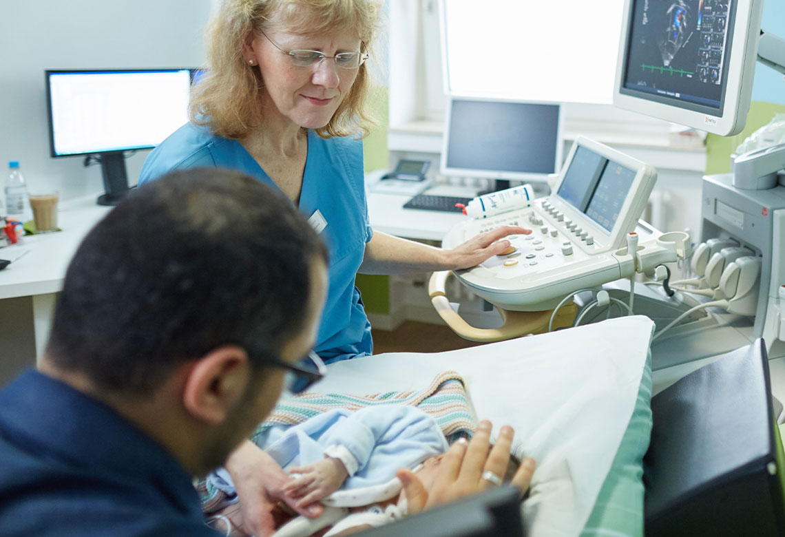
Ultrasound examination of the heart is the most frequently performed imaging method for examining the heart. New ultrasound probes and improved software enable high resolution and high frame rates. This method is therefore very well suited for diagnosing the heart with its rapid movements and fine structures - such as in the area of the heart valves.
Cross-sectional imaging procedures such as magnetic resonance imaging or computer tomography are further non-invasive imaging procedures that are used for a wide range of indications in cardiac diagnostics: MRI is a very good way of assessing the function and volumes of the heart cavities as they progress, completely without radiation exposure. The latest generation of computer tomographs allows excellent spatial resolution with minimal radiation dose.
More information on echocardiography More information on computed tomography More information on magnetic resonance imaging
Echocardiography
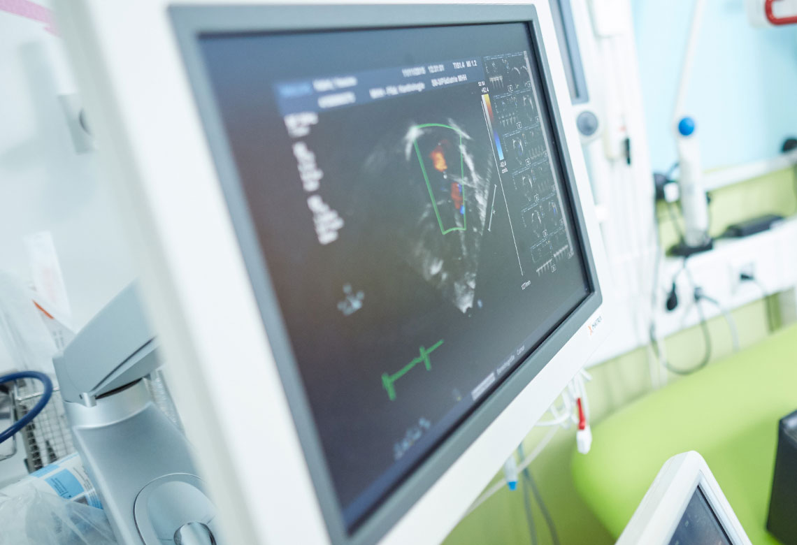
Transthoracic echocardiography is the basis for the diagnosis of congenital heart defects. The Department of Pediatric Cardiology and Intensive Care Medicine has all echocardiographic techniques at its disposal (2-D mode, M-mode, Doppler (PW, CW, color Doppler), tissue Doppler, 3-D echocardiography).
Precise functional analyses of the right and left ventricle can be carried out both globally and regionally by determining the myocardial velocity and deformity analyses (strain analyses). For this purpose, the tissue irregularities of the heart wall are tracked in their movement in the image data and the movement patterns are quantitatively evaluated ("speckle tracking").
In certain cases, the ultrasound examination is performed from the esophagus (TEE = transesophageal echocardiography). The patient "swallows" a tube with the ultrasound probe at the end (swallowing echo). Due to the proximity of the transducer to the heart and the good coupling to the heart, this examination makes it possible to accurately visualize fine structures of the heart, such as heart valves. TEE is now indispensable in the operating room. Before and during heart surgery, structures can be visualized in 3D to supplement surgical planning. The success of the operation can also be assessed in the operating room immediately after removal from the heart-lung machine. TEE is also regularly used during interventional cardiac catheter examinations, such as the closure of atrial septal defects (ASD), in order to be able to reliably close these defects in conjunction with the cardiac catheter.
Our Department has a total of two echo laboratories at its disposal, each equipped with high-end devices. One laboratory is integrated in cardiac ward 68 and one laboratory is available to the Department's outpatient clinics. In addition, a mobile high-end device is available in the intensive care unit and there is one device in the operating theater and one in the cardiac catheterization laboratory.
More information on our functional areas
Computed tomography
For patients with congenital heart defects, precise knowledge of the anatomy, in particular of scarring and coronary arteries, is extremely important.
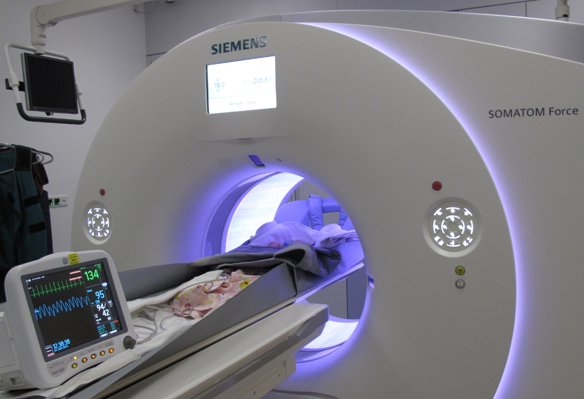
Since the beginning of 2016, Hannover Medical School has had one of the most modern CT systems available (Somatom Force, Siemens). With two radiation sources working simultaneously, the dual-source CT offers very high resolution in an extremely short time. After the administration of contrast medium via a venous vascular access, a complete scan is carried out in less than a second, showing all the vessels in the chest with very high resolution. Anesthesia is therefore not necessary for this very gentle examination, even for very small babies. Thanks to the detailed images, interventional cardiac catheter examinations and operations can be prepared precisely.
The use of dual-source technology further reduces radiation exposure compared to conventional CT systems. By integrating the 3D CT data sets obtained in this way into the planning software of the cardiac catheterization system, for example, the examination time in the cardiac catheterization laboratory can be reduced and the examination can be carried out with less radiation exposure.
More information on the integration of 3D data in the cardiac catheterization laboratory More information on the Institute of Diagnostic and Interventional Radiology
Magnetic resonance imaging
Magnetic resonance imaging (MRI) enables the anatomy and function of heart defects to be visualized precisely without the use of X-rays. This examination is therefore suitable for repeated use in the long-term course of patients with congenital heart defects. For example, changes in heart function (contractility) can be precisely quantified (volume-based determination of the ejection fraction, strain analyses) or flow phenomena on heart valves can be quantitatively assessed over time (flow velocities, regurgitation fraction, side-separated analysis of flows in the pulmonary vessels, estimation of collateral flows).
In the long-term course, this examination helps to determine the optimal time for an intervention - surgical or interventional - and also provides data on the anatomy.
With the help of "late enhancement" (accumulation of contrast medium over time in the heart muscle tissue), conclusions can be drawn about function, for example scar tissue can be identified. Inflammatory foci can also be localized in this way or indications of reduced blood flow can be obtained.
Our Clinical Department currently has access to a 3 Tesla MRI machine (Siemens), which we share with the Institute of Diagnostic and Interventional Radiology.