During your stay at the Clinical Department of Paediatric Cardiology and Intensive Care Medicine, you or your child will get to know one or more of the functional areas. The functional areas ensure that the necessary examinations and interventions are carried out smoothly and are briefly introduced below.
The transthoracic ultrasound examination of the heart, i.e. the examination from the outside through the chest, is a painless examination. Thanks to the ever-improving image quality, many diagnostic questions can now be answered with the echo alone. Valuable information on the structure and anatomy of the heart as well as its function can be obtained using this method. Our ultrasound devices support the creation of 3D images in real time. Software analysis of wall movements (strain analysis) allows statements on function to be quantified. In addition to the ultrasound examination from the outside through the chest, additional information can be obtained by an examination through the esophagus (TEE, transesophageal echocardiography).
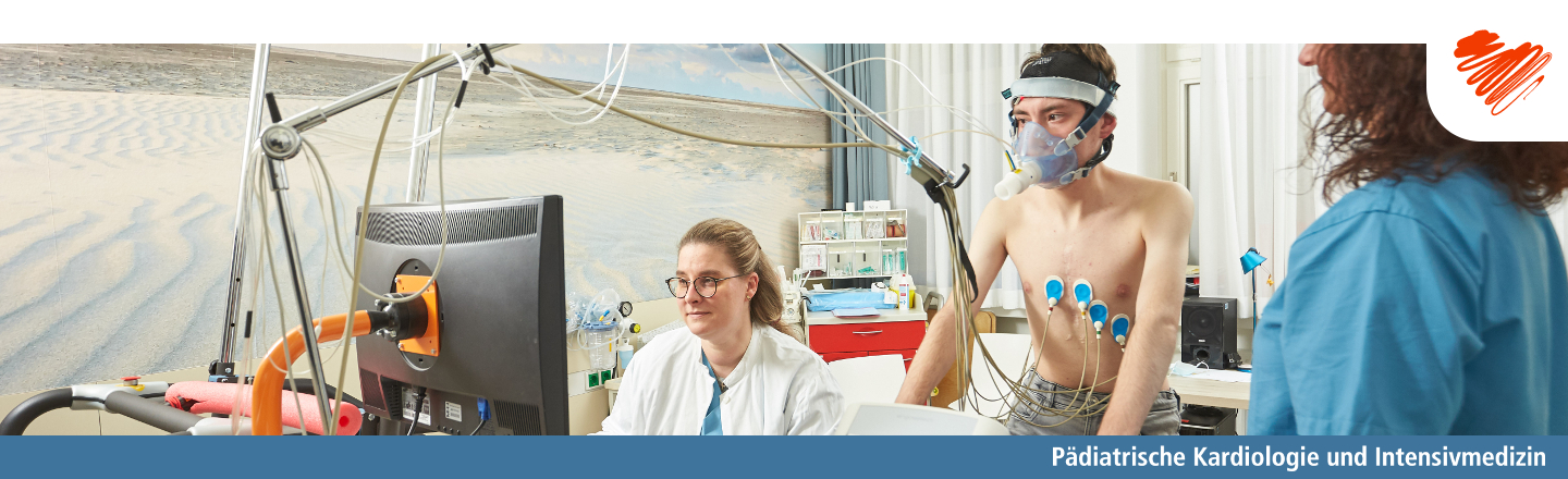
Cardiac performance diagnostics are carried out either on the treadmill or on the bicycle ergometer. To measure oxygen uptake, a mask is worn over the mouth and nose during the examination. The exertion is achieved over time by increasing the resistance on the cycle ergometer or by increasing the speed and incline on the treadmill. The test ends when the load limit is reached. Blood is taken from the finger to determine the lactate (metabolic product in the case of an anerobic metabolic state) in order to assess the level of exertion. The performance test can help to determine when to take further measures.
The recording of cardiac currents is a painless and uncomplicated way of obtaining information about the heart rhythm. However, the excitation processes of the heart can also be used to draw conclusions about the course of diseases of the heart muscle and the effects and side effects of drug therapies.
In addition to the recording of a directly derived ECG, it is sometimes necessary to record over a longer period of time (e.g. 24 hours). For this purpose, a small device is connected to electrodes on the body and continuously records the heartbeat. With a small neck strap, it can also be carried during light "sporting" activities such as running or climbing stairs. However, it must not come into contact with water (no showering, bathing or swimming). If the heart rhythm needs to be monitored over a longer period of time, other systems - such as an event recorder - are used.
The Medical School has one of the most powerful dual-source computed tomography (CT) scanners currently available. With two radiation sources and two detectors, the complete anatomy of the heart and blood vessels can be imaged within half a second, with a very low dose of radiation. This high-resolution image allows precise planning of interventions or surgical measures. The examination does not require anesthesia or respiratory arrest and can therefore also be performed on very small infants.
Computed tomography is carried out in cooperation with the Clinical Department of Radiology at Hannover Medical School.
Magnetic resonance imaging (MRI) examinations of the heart provide information on anatomy, function and tissue properties. The examination is painless, but requires the patient to follow breathing commands (e.g. "hold your breath"). As this does not always work with small children, MRI examinations are occasionally carried out under anesthesia, but this is often not necessary.
The examination is based on the evaluation of certain tissue properties under the influence of strong magnetic fields. The extremely fast switching of the magnetic fields results in loud noises; however, the examination is neither dangerous nor associated with radiation.
The MRI examinations are carried out and analyzed in cooperation with the Clinical Department of Radiology at Hannover Medical School. They allow an assessment of, for example, the indication and urgency of further measures such as surgery or cardiac catheter therapy and help with the planning of these measures.
The cardiac catheterization laboratory in the Pediatric Cardiology Department at Hannover Medical School is one of the most modern laboratories in Europe. Using low-radiation technology, the entire spectrum of therapies and diagnostic examinations can be offered here.
In most cases, an access in the groin is required for the examinations and the patient must lie quietly on their back during the examination. The examinations are therefore often carried out under sedation. This means that the patient is given medication that puts them to sleep and makes them pain-free - deep sedation "feels" the same to the patient as anesthesia, they are not in pain.
Parents can accompany their child into the examination room and - if they wish - stay with them until they fall asleep.
The operating theater - actually operating room - at Hannover Medical School is equipped with state-of-the-art cardiotechnology. Cardiotechnology ensures that the body is not damaged during operations on the heart and the vessels close to the heart and that all organs are supplied with sufficient oxygen.
The congenital heart surgery team from the Department of Cardiothoracic and Vascular Surgery can perform even the most technically difficult operations in the operating theater under optimal conditions and offers the entire spectrum of surgical care.
On the day of the operation, parents can accompany their child until shortly before the operation. There, they are welcomed by anesthesia colleagues and often given medication to help them fall asleep before they are separated from their parents.
Once the heart operation has been completed, the patient is transferred to the intensive care unit for further care and monitoring.