Cardio-pulmo-vascular radiology team
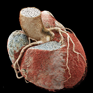
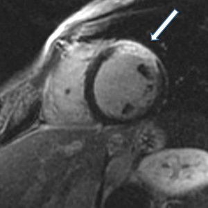
Cardio-pulmo-vascular radiology deals with the thoracic organs (heart, lungs) and adjacent structures such as the aorta and pulmonary arteries.
Contact persons: Prof. Dr. D. Hartung, Prof. Dr. J. Vogel-Claussen
Procedure: We use the following procedures to examine pathological changes in the thoracic organs, such as inflammations and tumors of the lungs, circulatory disorders, inflammations or other diseases of the heart muscle, diseases of the heart valves, congenital heart defects, dilatations, constrictions or injuries of the aorta and its outgoing vessels or occlusions of pulmonary arteries: