PERCUTANEOUS TUMOR ABLATION
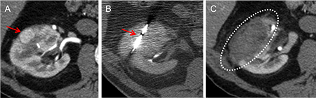
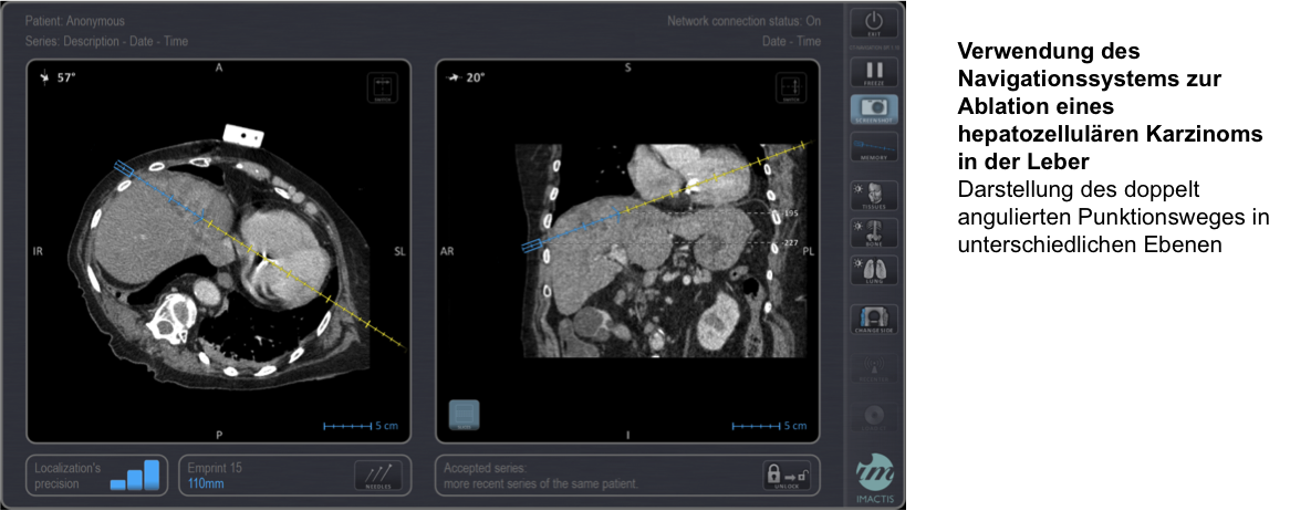
Percutaneous tumor ablation has a long tradition at the MHH and has been successfully performed here for over 15 years. These are non-surgical image-guided therapy procedures for the treatment of benign and malignant tumors, e.g. of the liver, kidneys, lungs or bone. Under computer tomography (CT), magnetic resonance imaging (MRI) or ultrasound control, special probes are placed directly in the tumor and, depending on the procedure, heat (hyperthermic ablation) or cold (cryoablation) is generated. This temperature change in the tissue leads to the destruction of the tumor cells, which are broken down over time by the body's own immune system.
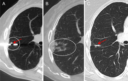
Various techniques are available. At our Institute, we use in particular the two most widely used and best evaluated procedures in clinical studies, in the form of radiofrequency (RFA) and microwave ablation (MWA). In RFA , one or more probes are heated to 70-90° Celsius for 5-60 minutes, depending on the size and number of tumors, by directly applying electrical energy. In MWA, the antennas are also placed directly in the tumor. In this procedure, the heat is generated indirectly through the interaction of the electromagnetic radiation with water molecules in the tissue and can reach temperatures of over 150° Celsius. Each method has certain advantages and disadvantages, and the choice ultimately depends on the size, number, exact location and type of tumor, among other things.
The decision as to whether ablation is an option for the patient is made at Hannover Medical School together with colleagues from other specialist departments (surgery, oncology, gastroenterology) as part of a tumor conference. This ensures that the optimal treatment procedure is chosen, taking into account all alternatives and any concomitant therapies.
The procedure usually only requires a short hospital stay of two to three days. The day before the intervention, a detailed information session is held and the blood values are checked. If necessary, up-to-date images (CT, MRI or ultrasound) are also taken for precise planning. The procedure is performed on the second day, usually under general anesthesia. After a short period of monitoring in the recovery room and on the ward, the patient is usually discharged the following day.
Contact / contact person:
If you would like to know whether tumor treatment is possible for you or a friend, for example, or if you would like further information, please contact us or make an appointment for a consultation.
Hannover Medical School
Institute of Diagnostic and Interventional Radiology
Director: Prof. Dr. med. F. Wacker
Office: Phone +49 (511) 532 3421
Contact for tumor ablation: PD Dr. med. K.I. Ringe