AG Bone / Fracture Healing
Fractures are still not uncommon these days. Normally these heal without complications or scarring. However, if a fracture has not completely bridged the bone after four months, a fracture healing disorder or delayed union is present. If there is still no stable bridging after six months, this is referred to as pseudarthrosis ("non-union"). There are many reasons for delayed or non-union. The most common causes are
- inadequate blood supply, e.g. due to a lack of vascularization, resulting in bone necrosis,
- insufficient stabilization of the fracture, so that there is too much mobility in the fracture gap,
- a lack of contact between the bone fragments
- or comorbidities such as infections, age, alcohol consumption, smoking, osteoporosis or polytrauma.
The "Bone/Fracture Healing" working group is therefore investigating various possible causes of fracture healing disorders and pseudarthrosis. As it is difficult to standardize and obtain bone samples in a clinical context, animal models are often used. However, the overarching goal is always to better understand the underlying mechanisms of fracture healing disorders in order to identify patients at risk at an early stage and develop new therapies.
Fractures are often associated with severe blood loss due to vascular injury, and clinical studies show that fracture healing can be impaired and delayed in patients who have also suffered severe blood loss. Therefore, investigating the effect of such severe blood loss on fracture healing is a particularly important topic.
In the mouse model, our research group has already been able to show that severe blood loss has a negative effect on bone regeneration after fracture in young healthy mice. This has been demonstrated both macroscopically and in X-ray images and µCT scans. The biomechanics of the bones also showed a reduced maximum bending moment in the 3-point bending test and histologically less bone and cartilage with severe blood loss was detectable in the fracture gap compared to an isolated fracture. Furthermore, using PCR arrays, qRT-PCR with TaqMan assays and Western blot analysis, we found that the Wnt signaling pathway is strongly affected by severe blood loss. This is important for bone metabolism and is involved in both bone formation and resorption, so that inhibition also led to delayed bone healing in this model. However, since the average patient is not young and is often under the influence of alcohol in addition to the trauma, this mouse model of severe blood loss in combination with a fracture was also used under the influence of alcohol and/or in combination with old age.
If you have any questions or suggestions, or if you are interested in writing a master's or doctoral thesis in our laboratory, please contact us!
Tel: +49 511 532-84246
Bundkirchen.Katrin@mh-hannover.de
Fracture healing in the mouse
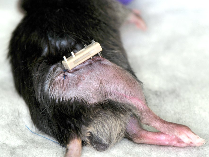
An established model in fracture healing research that we also use is the stabilization of a bone fracture using an external fixator, which is also used in the human Clinic.
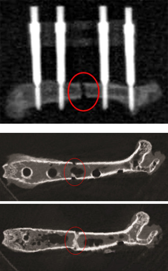
Radiological evaluation
A) X-ray image of a previously fractured bone including an inserted external fixator.
B) µCT scans after removal of the external fixation. The red circle marks the fracture.
Histological evaluation
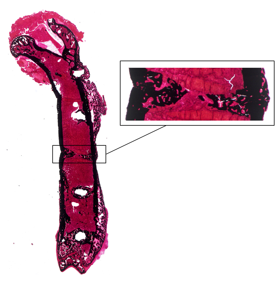
The mineralized bone in the fracture gap can be evaluated using Kossa/Safranin O staining. This is shown in black, while all other tissue types such as bone marrow, connective tissue and muscles are stained in different shades of red.
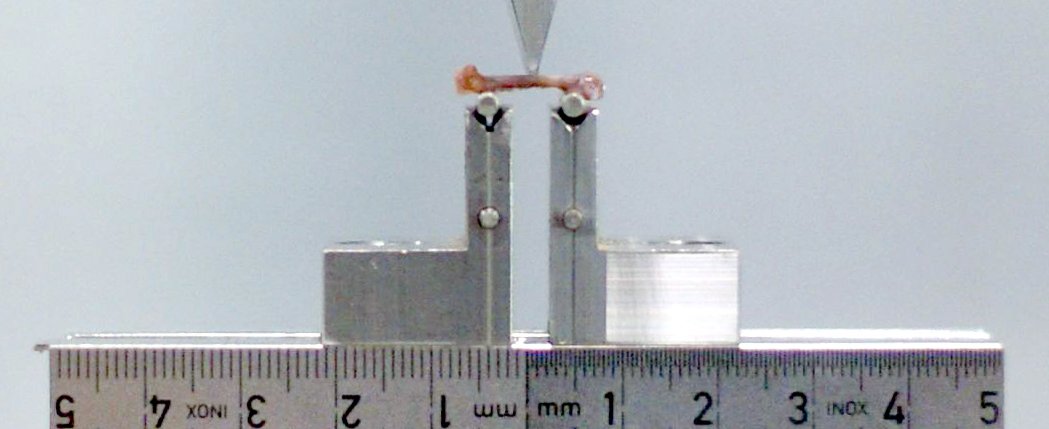
Biomechanical evaluation
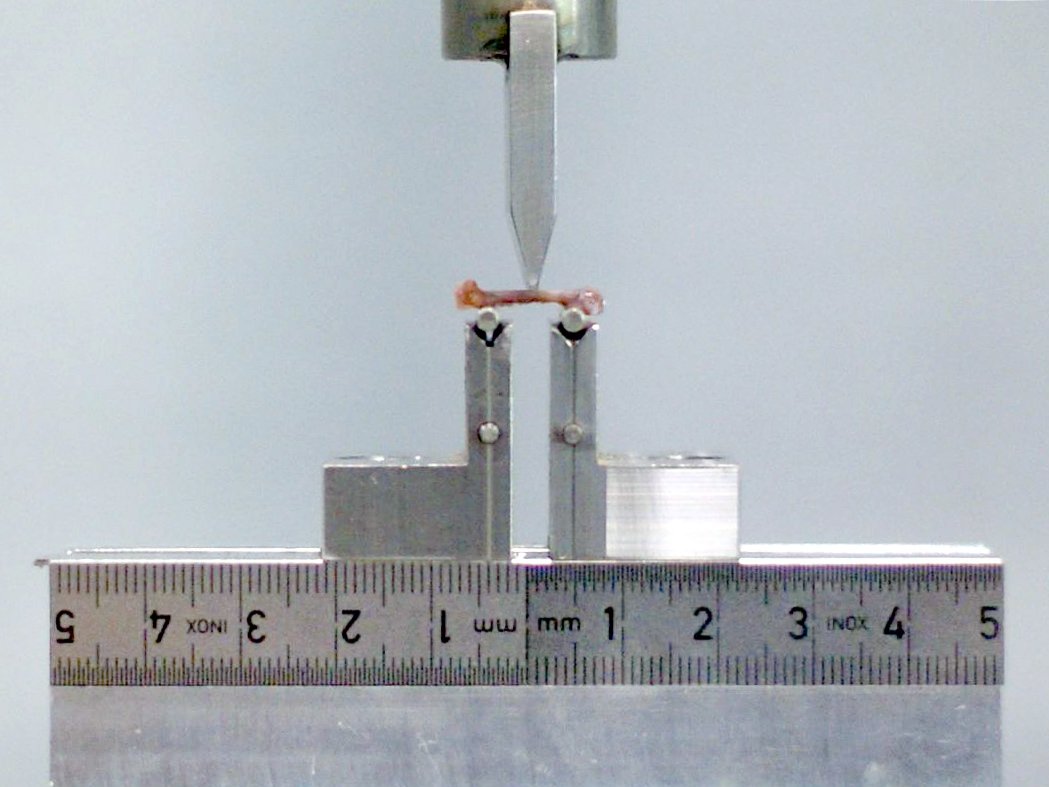
The bones are analyzed using the 3-point bending test. To do this, they are placed in anteroposterior alignment on a specially designed apparatus. A punch presses on the sample from above until structural failure occurs.