to the Interdisciplinary Tumor Conference
of the Comprehensive Cancer Center of the MHH
Online study for sarcoma patientsto "Evaluation of the rehabilitation of sarcoma patients and their provision with aids and remedies - REHSa"
The early detection and stage-appropriate surgical treatment of joint and skeletal tumors has a long tradition as a focus in the Clinical Department of Trauma Surgery. Patients with unclear processes or tumors of the musculoskeletal system receive targeted diagnostics and detailed advice in the special clinic for joint and skeletal tumors. We understand the emotionally upsetting situation in the phase of diagnostic uncertainty and will contribute to a rapid diagnosis with suitable imaging and the initiation of subsequent medical measures, such as taking tissue samples.
Our interdisciplinary tumor conference for bone and soft tissue tumors was founded in 1979 as one of the first tumor conferences at the MHH.
If you have a tumor diagnosis relating to the musculoskeletal system that requires clarification, you are welcome to present yourself at the special clinic for joint and skeletal tumors.
Based on the findings compiled in the consultation, your case will be discussed in the tumor conference for bone and soft tissue tumors if interdisciplinary consultation is required, and the appropriate treatment concept will be determined for you individually.
Examination, treatment & aftercare
Local tumor spread
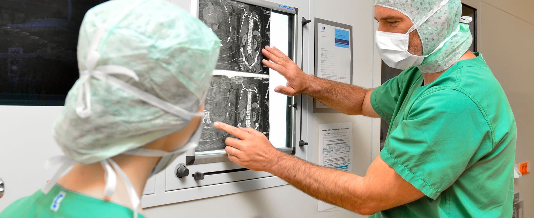
- X-ray image in 2 planes
- Ultrasound diagnostics (sonography)
- Magnetic resonance imaging (MRI with contrast medium)
- Computed tomography (CT with contrast medium)
Body-wide tumor spread (so-called staging)
- Computed tomography of the chest and abdomen (CT thorax & abdomen)
- 3-phase skeletal scintigraphy (bone scinti)
- Positron emission tomography (PET CT)
A definitive tumor diagnosis can often only be made after a tissue biopsy has been performed. If a malignant tumor is still suspected after imaging, a biopsy is absolutely necessary. In the case of soft tissue tumors, a tissue sample can usually be obtained minimally invasively using a punch biopsy (hollow chamber needle) after interpretation of the MRI imaging. In the case of primary bone tumors with hard basic substance of the tumor, we usually perform an incisional biopsy (open biopsy) under general anesthesia under inpatient conditions. A small "preliminary operation" is therefore necessary. This should be carried out in a center with experience in the treatment of tumors of the musculoskeletal system. Experience has shown that an incorrect biopsy technique can make definitive tumor removal significantly more difficult or increase the risk of local recurrence. We pay attention to meticulous hemostasis and avoid unnecessary contamination of healthy tissue.
Depending on the type of tumor, tumor removal follows basic surgical principles (Enneking criteria). Important for surgical therapy is the definition of the border zone between the tumor tissue and the surrounding healthy tissue.
- Intralesional resection means partial removal of the tumor from the inside, i.e. the tumor is opened and removed in several parts.
- Marginal resection means surgical removal of the tumor as a whole (en bloc) without opening it but close to the tumor pseudocapsule
- Wide resection means complete surgical removal of the tumor and the pseudocapsule with a sufficient safety margin (healthy tissue cuff), taking the biopsy area with it.
- Radical resection means complete surgical removal including the tumor-bearing tissue/muscle layer, accepting severe functional deficits.
While benign tumors can usually be adequately removed by marginal resection, a wide or even radical resection should be attempted in the case of malignant bone and soft tissue sarcomas. The most unfavorable margin to the tumor, however small it may be, is decisive for the prognostic assessment. Thus, local elimination of the tumor has the highest priority, and preserving the function of a limb remains secondary to the primary goal.
Remaining bony defects after tumor removal require reconstruction of the affected skeletal section.
Biological reconstruction
Autologous bone transplantation (autograft)
- Cancellous bone
- Bone chip
- Vascularized fibula
Foreign bone transplantation (allograft)
- Femoral heads
- Bone granules
Bone transportation
- Callus distraction
- Segment transport
Non-biological (alloplastic) reconstruction
Tumor endoprosthetics (megaprostheses, e.g. MUTARS)
- Alloplastic joint replacement (hip, knee, shoulder and elbow)
- Alloplastic skeletal replacement (pelvis, thigh, tibia, upper arm)
Endoprosthetics with modular megaprosthesis systems have a firm place in the treatment of bony defects following tumor removal. Almost any skeletal segment can be replaced thanks to the variability of the prosthesis systems.
Important information
Interdisciplinary tumor conference for bone and soft tissue tumors
Mondays 16:00 - 17:30
in the radiology lecture hall (S level, node E)
Registration
by 12.00 noon on the Thursday of the previous week
Incl. imaging on CD and referral
for the attention of Ms. Maren Herten
Phone: +49 511 532-5818
Fax: +49 511 532-5877
uch-team-tumor@mh-hannover.de
Head
N.N.
As a basis for a differentiated consultation, please bring the following documents with you to your appointment:
- Referral from a specialist in surgery, trauma surgery or orthopaedics
- Previous imaging (X-ray/CT/MRI) on a CD ROM in DICOM format, incl. findings/diagnoses.
- Reports from previous treatments (e.g. surgical reports)
- Reports of tissue samples (pathology findings)