Our team for shoulder and elbow surgery at Hannover Medical School deals with all injuries and conditions of the shoulder and elbow joint. We treat patients both in emergencies and chronic diseases, or in the event of treatment failure.
Another focus of activity is research and development in cooperation with numerous doctoral students, scientific staff and in cooperation with renowned research partners and specialist surgical societies. Research results are regularly published and discussed at congresses and in journals. As part of the MHH teaching staff, key aspects and innovations in the field of shoulder and elbow surgery are taught to medical students in lectures, internships and courses. In addition, regular further training of medical colleagues from various specialties takes place, for example as part of professional events and work shadowing.
We offer patients and colleagues specialized consultations once a week. In order to optimise workflows and waiting times, we ask you to bring all existing diagnostics (X-rays, CT, MRI, findings and reports) with you. Due to the high number of patients and the complexity of some cases, there may be delays on the day of the appointment. We apologize for this in advance.
In emergencies, patients can be seen at any time via the MHH Central Emergency Room (ZNA).
Please contact us if you have any questions!
Main areas of treatment
Introduction
In contrast to other joints, the shoulder joint is largely stabilized by capsule, muscle and tendon structures (tendon cap). Accidents can lead to fractures in the area of the shoulder joint and the adjacent bones, but also to tears in the tendon and capsule structures. On the other hand, age-related changes to structures of the shoulder joint can lead to painful processes and similar clinical pictures. A combination of both entities is also not uncommon.
Shoulder surgery has developed enormously in recent decades with new techniques and minimally invasive procedures. Many shoulder operations can be performed arthroscopically via small skin incisions.
A precise and targeted medical history and diagnosis is essential for the treatment decision and therefore forms the basis for successful treatment. The treatment decision is based on the assessment of both the bony and tendinous changes in the shoulder joint.
Definition and diagnosis
A dislocation of a joint occurs when the socket and head of the joint are no longer in contact. This usually occurs as a result of an accident. As a rule, a reduction by a physician is required. In rare cases, the patient can reduce the shoulder themselves. If the joint is not completely dislocated, it is referred to as a subluxation. Typically, the labrum, the capsular ligament structures and the humeral head are injured. In rare cases, there may be tears of the rotator cuff, fractures of the tuberosity, or nerve injuries.
An accident-related (traumatic) dislocation of the shoulder joint is painful and associated with a temporary loss of shoulder function. Immediate pain relief occurs after reduction. Depending on defined parameters, healing may subsequently occur without further symptoms. In other cases, symptoms may develop that make surgery necessary.
Acute diagnostics prior to reduction include x-rays of the shoulder. The X-rays are repeated to check the reduction. An ultrasound examination is performed to rule out a rotator cuff tear. Further diagnostics (MRI, CT) depend on the patient's risk profile.
Therapy
Conservative
After creating an individual profile, the risk of recurrent dislocation is calculated. Patients with a low risk profile are treated by immobilization for three weeks and subsequent physiotherapy.
Surgical
patients with a high probability of re-dislocating are advised to undergo surgery. Traditional surgical treatment involves refixation of the injured labrum and capsular ligament structures. Transfer surgery of the coracoid (Latarjet surgery) is increasingly being considered.
Follow-up treatment and results
Immediately afterwards, the shoulder is immobilized in an orthosis. Physiotherapeutic exercise already begins during this phase. Full weight-bearing capacity of the shoulder is normally restored in 6 to 12 weeks.
Diagnosis
This is an injury to the four tendons that surround and attach to the humeral head. Tears to this tendon cap caused by an accident must be distinguished from degenerative tears caused by wear and tear in old age. However, there are often mixed forms, i.e. acute tears caused by an accident with a degenerative, pre-damaged rotator cuff. The biceps tendon may be injured in other structures and, in rare cases, intra-articular structures such as the labrum or articular cartilage.
A rupture of a rotator cuff tendon is usually described by patients as a painful event. A corresponding accident mechanism could be a fall on the shoulder or a lifting trauma. This usually results in an acute pain situation with a restriction of movement, but above all a loss of strength for certain movements.
In the acute phase, standardized x-rays of the shoulder are routinely performed in 3 planes. Clinical tests provide an initial indication of an injury to the rotator cuff. Sonography of the shoulder joint is also performed. To confirm the diagnosis, magnetic resonance imaging and possibly computer tomography with contrast medium are required.
Therapy
Conservative
tears in the case of pre-existing tendon wear, in patients of advanced age, in patients with concomitant diseases, and partial tears of the rotator cuff can initially be treated conservatively.
Surgical
Patients of working age with a documented rupture of the rotator cuff should undergo surgical refixation. Ruptures of the subscapularis tendon in particular are considered an orthopaedic emergency and should be treated promptly. The procedure of choice is arthroscopic, i.e. minimally invasive, refixation of the torn tendon using special anchoring techniques.
In the case of pronounced and extensive tears of the tendinous shoulder cap, it may no longer be possible to reconstruct the tendon. In these cases, special techniques are used such as balloons that are inserted arthroscopically below the acromion, or special reconstruction techniques of the tendon using artificial skin or muscle transfers.
Follow-up treatment and results
Conservative treatment usually leads to a continuation of the rupture, a so-called "spontaneous healing" without surgery is described in the literature, but is only observed in partial ruptures. After the tendon has been reattached, the shoulder joint is immobilized in a special orthosis to ensure that the tendon heals. Physiotherapeutic exercise in a pain-free environment can be started at an early stage. In the majority of patients, a mostly pain-free situation can be achieved.
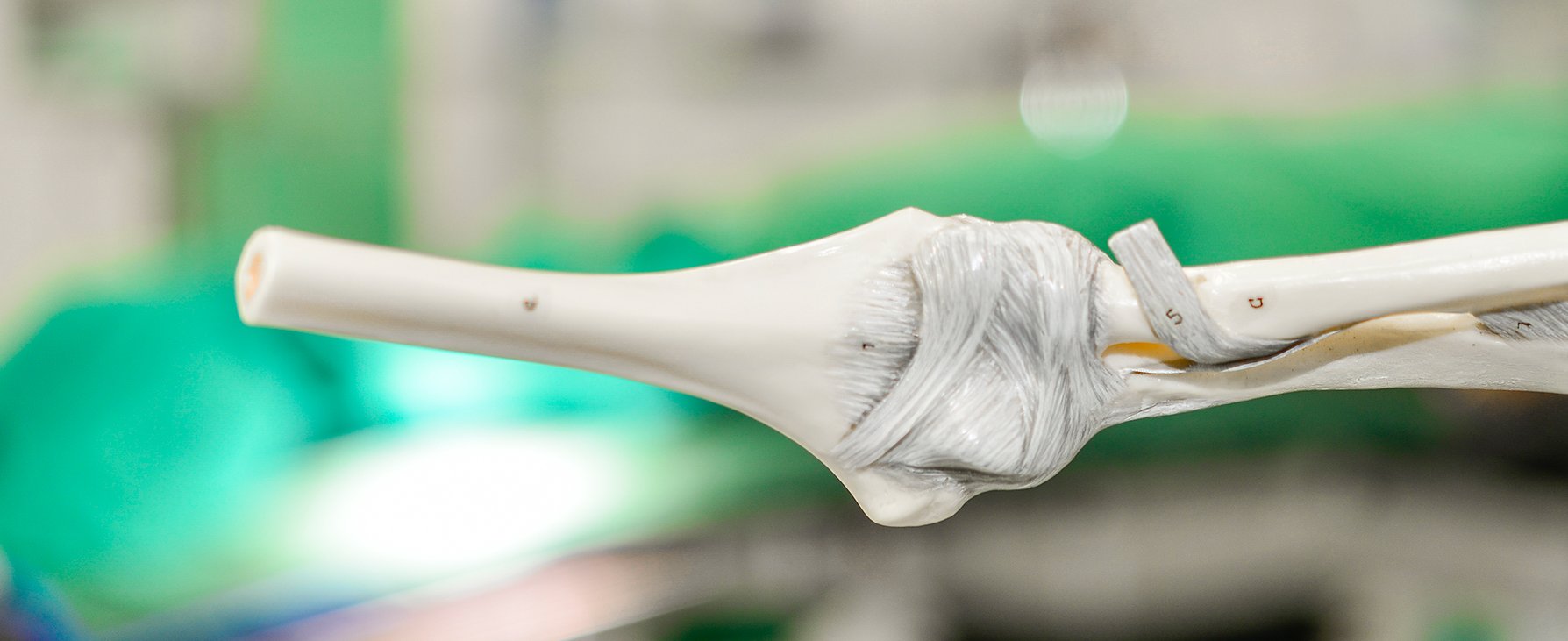
Diagnosis
The biceps muscle of the upper arm consists of two heads with two muscle insertions at the end close to the body. The tendon of the long biceps muscle originates inside the shoulder joint at the upper edge of the glenoid cavity. The tendon runs in the joint under the tendon cap into a suspension system (pulley system) and then away from the body on the humerus. The humeral head therefore slides directly underneath the tendon of the long biceps muscle. Both age-related processes and accidents can lead to inflammation, (partial) ruptures and changes at the origin of the tendon. Furthermore, the suspension system can be altered and torn due to age or accident, which can lead to injuries to the adjacent structures, and the tendon can also pop out of the bearing.
Therapy
Conservative
Minor severity inflammation of the long biceps tendon can be treated conservatively. A combination of oral anti-inflammatory drugs and physiotherapy is usually sufficient.
Surgical
More severe or treatment-resistant injuries should be addressed surgically due to the possible long-term consequences. Patients can be treated entirely arthroscopically. Depending on the intraoperative findings, the tendon can be fixed to the humeral head using an anchor (tenodesis) or simply severed at its origin (tenotomy). In the latter case, the muscle belly may deepen. If the tendon becomes loose or frayed at its origin, it can be smoothed electrothermally or fixed with anchors.
Aftercare and results
After the operation, the upper arm is immobilized in a sling. Depending on the operation, you should refrain from exerting the biceps muscle. Physiotherapeutic exercise can be started early on, but this should only ever be carried out to the pain threshold. Complete recovery of shoulder function can be expected after 6 to 12 weeks.
Diagnosis
Impingement syndromes are clinical pictures in which constrictions occur between the humeral head and the acromion or coracoid (coracoid process). This impingement causes the tendons of the shoulder cap and the adjacent bursa to become trapped. Patients often report night pain and pain on movement. There may also be a loss of strength. The clinical examination provides the first indication. Bony changes are detected on the basis of the X-ray image. An MRI examination is the best way to visualize the narrowing of the tendon cap.
Therapy
Conservative
Conservative treatment should be initiated first, with temporary rest, anti-inflammatory medication, local ice/heat applications and physiotherapy exercises. In this context, incorrect posture of the scapula, which can also lead to this bottleneck (scapular dyskinesia), can also be corrected.
Surgery
If conservative therapy is unsuccessful, arthroscopy can be used to gently widen the existing bony constriction. In addition, any pathology present (tendon already damaged by the bottleneck) can be addressed therapeutically.
Follow-up treatment and results
The shoulder joint is only immobilized immediately postoperatively. Physiotherapeutic exercise in a pain-free environment can be started at an early stage. There is usually a reduction in pain within 6 to 12 weeks.
Diagnosis
Calcium deposits can occur in the area of the tendon cap of the shoulder. This calcium deposit is located inside the tendon and can be very painful intermittently. Patients usually complain of night pain and acute restriction of movement.
After the clinical examination, the main diagnostic tool is the X-ray. This is where the calcium deposit is detected and localized. MRI imaging can reveal additional pathologies.
Therapy
Conservative
The progression of a calcified shoulder is phased until spontaneous resorption and thus healing. However, the time course cannot be calculated. Conservative approaches include shock wave therapy or needling.
Surgical
If pain persists and conservative treatment does not work, surgical removal of the calcium deposit is recommended. The calcium deposit is cleared out and removed arthroscopically via small skin incisions. A small part of the calcium deposit often remains, but this resorbs by itself over time. Arthroscopy allows other pathologies in the shoulder to be diagnosed and treated if necessary.
Follow-up treatment and results
Immobilization is only required immediately postoperatively. Physiotherapeutic exercise begins early after the operation. Medication to bring down swelling can provide post-operative support. In most cases, a pain-free situation can be achieved.
Diagnosis
This is characterized by inflammation of the joint capsule. Patients report severe pain with even slight movement, which is often described as knife-like. Mobility is only restricted as the condition progresses. The clinical picture of frozen shoulder is self-limiting up to spontaneous healing, but the course over time cannot be calculated.
Therapy
Conservative
In the early stages, treatment involves taking cortisone according to a certain regimen. Once the symptoms have subsided, pain-adapted physiotherapy can be started.
Surgical
If the pain and restriction of movement do not improve despite conservative therapy, the joint capsule can be opened and the inflamed tissue removed by means of arthroscopy. This can significantly shorten the course of the disease and improve the final result.
Follow-up treatment and results
Immobilization only takes place immediately after the operation. Patients are given a pain catheter during the operation, which is used for pain therapy after the operation. Intensive physiotherapy and self-exercises are started immediately after the operation.
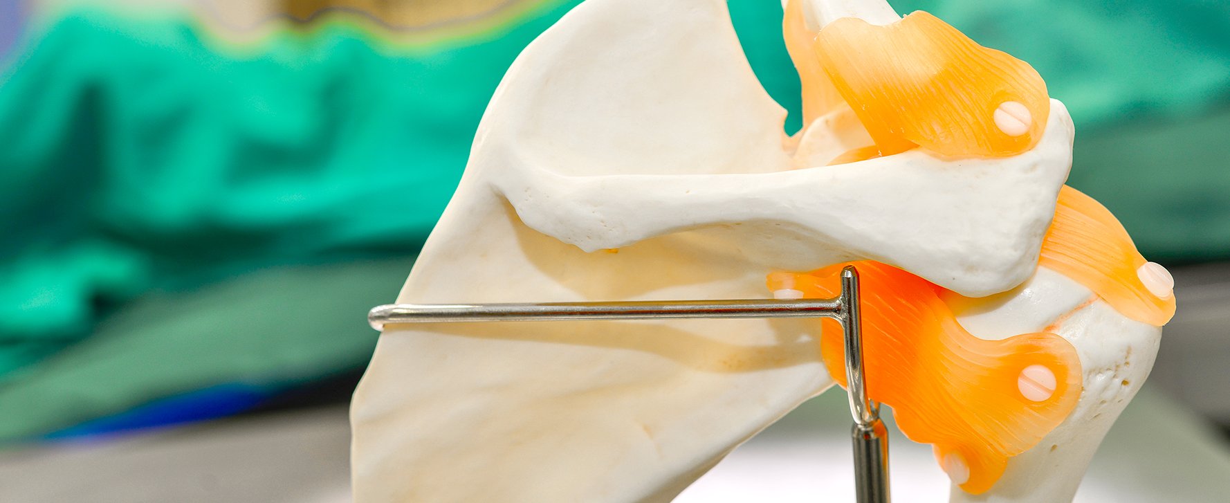
Diagnosis
The acromioclavicular joint is located between the acromion and the clavicle. The distal end of the clavicle is connected by ligaments to both the acromion and the scapula (coracoid, coracoid process) and thus stabilizes the acromioclavicular joint. Typically, a fall on the shoulder can lead to these ligaments being torn. Patients show a painful restriction of movement and pain in this area. Visually, depending on the number of ligaments affected, the clavicle is elevated above the shoulder (piano key phenomenon). Manual pressure on the clavicle also triggers pain. In addition to the medical history and clinical examination, an X-ray shows the extent of the injury in a lateral comparison.
Therapy
Conservative
Up to a certain degree (Rockwood type III), there is no indication for a surgical procedure. In this case, immobilization and temporary avoidance of weight-bearing and abduction movements above the horizontal are recommended.
Surgical
If the ligaments are severely torn and the clavicle is clearly elevated, arthroscopically assisted rein can be performed. Special sutures are passed through the part of the clavicle remote from the body and the coracoid. Once the congruence of the acromioclavicular joint has been restored, these sutures are then tied above the clavicle. In the case of an older injury, a transfer of the patient's own tendons is required to ensure healing.
Follow-up treatment and results
After surgical stabilization, immobilization is required to allow healing to take place and to prevent the stitches from pulling out. After around 6 to 8 weeks, the range of motion can be extended in a pain-adapted manner. Patients usually achieve pain-free mobility after 3 to 6 months.
Diagnosis
The acromioclavicular joint is located between the acromion and the clavicle. Like every joint, the acromioclavicular joint is subject to signs of wear and tear with wear of the joint cartilage and bony changes. Patients report localized pain, especially during movement and exertion. Lying on the side can also be very uncomfortable.
Therapy
Conservative
If there are no or only minor symptoms, no treatment is required. If necessary, pain relief can be achieved with a local injection.
Surgical
If the symptoms persist over a longer period of time, surgery is possible. Arthroscopy can be used to remove a small part of the collarbone above the shoulder. This prevents constant rubbing and chronic irritation of the joint.
Aftercare and results
Immobilization is only required immediately after the operation. Pain-adapted physiotherapy can be started at an early stage. There is usually a reduction in pain within 6 to 12 weeks.
Diagnosis
Age-related signs of wear and tear, certain diseases or accidents can lead to a loss of cartilage and destruction of the shoulder joint. Osteoarthrosis can affect the entire joint or only a limited area. This clinical picture is usually accompanied by severe pain and restricted movement. The medical history and clinical examination provide the first indication. Conventional X-rays and an MRI or CT scan are also used for diagnosis.
Therapy
Conservative
The early, low-pain phase of osteoarthrosis can initially be treated conservatively with physiotherapy and local application of heat or ice. It is important to protect the joint and avoid overloading it.
Surgery
In the case of less pronounced osteoarthrosis, an arthroscopy can be performed if the condition is painful. Inflammatory tissue is removed and the cartilage is smoothed. Many patients achieve a significant improvement in their symptoms as a result. Even if this measure does not halt the progression of osteoarthrosis, it is a much gentler procedure than joint replacement surgery.
If the cartilage defect is limited to a specific area of the humeral head, this specific part of the humeral head can be replaced arthroscopically and without tendon detachment. The advantage of this method is that the patient retains the majority of their intact humeral head. This procedure is mainly used in young patients with localized cartilage damage.
With increasing loss of function and advanced osteoarthrosis of the entire joint, a joint replacement is performed. If the cartilage of the joint socket is still intact, it may be sufficient to replace only the humeral head with a prosthesis. The humeral head prosthesis has a crescent-shaped appearance with a short thread or short stem, which is anchored in the humeral head to save bone. If the joint socket is also damaged, this must also be replaced by an artificial surface.
In cases where the tendon cap of the shoulder joint has already been destroyed and osteoarthrosis is also present, special prostheses are used. A hemisphere is implanted in the socket and a shell in the humeral head. This practically reverses the joint and makes it possible to use the remaining muscles to move the shoulder joint.
Aftercare and results
Both postoperative immobilization and follow-up treatment depend on the form of therapy and the chosen prosthesis. Physiotherapy treatment is required over a period of 3 to 6 months. A prosthetic restoration leads to pain-free function in the majority of patients. The range of movement after the operation depends on the functional capacity before the operation.
Diagnosis
These are fractures of the upper arm in the shoulder area. These fractures typically occur at an advanced age of 65 years or older, usually as a result of falls on the upper arm. They are often associated with reduced bone density (osteoporosis). Women are more frequently affected than men. In addition to injuries to the skin and soft tissue mantle, which together with the fracture are referred to as open fractures, fractures of the socket of the humeral head and dislocations of the shoulder can occur in rare cases.
Fractures of the humeral head are usually very painful and initially lead to a loss of function of the shoulder and arm. In rare individual cases with severe malalignment of the fracture, the blood circulation and sensitivity of the arm may also be impaired, as the main vessels and nerves of the arm run in the immediate vicinity of the humeral head.
X-rays of the shoulder are taken in the acute phase. A computer tomography may be necessary to decide on further treatment, e.g. with regard to surgery.
Therapy
Conservative
Non-displaced or slightly displaced 2- and 3-part fractures, submerged fractures of type A2.2 and B1.1 of the AO classification and submerged 3-part fractures without a twisted head fragment can be healed without surgical treatment. Pronounced osteoporosis and serious concomitant diseases with a high risk of anaesthesia complications are further criteria for conservative treatment. The first step is immobilization in a Gilchrist bandage. After 1 week, the first pendulum exercises are started. This is followed by a graduated treatment regimen with passive and active physiotherapy. From the 6th week, movement of the shoulder is generally allowed. After approx. 3 to 5 months, the level of activity as before the accident is reached again.
Surgical
Severely displaced 4-part fractures, dislocated fractures regardless of age, head gap fractures and open fractures require surgical treatment. A secondary dislocation of the humeral head fracture after initial conservative treatment may also require surgery. Depending on the fracture morphology, concomitant diseases, patient age and functionality, either a plate is attached to the side of the humeral head. However, it is also possible to replace the humeral head and thus the shoulder joint with an endoprosthesis, similar to the knee or hip.
Aftercare and results
According to current knowledge, the vast majority of humeral head fractures (approx. 70-80%) can be treated conservatively without surgery. Current studies show that patients do not always benefit from surgical treatment and that revision surgery is often necessary. It is therefore important to consider the patient's risk profile.
Timely initiation of physiotherapy and good patient guidance and cooperation play an important role in the final outcome. Restrictions, particularly for movements in the end range of shoulder mobility and overhead work, as well as loss of strength are often not subjectively perceived as restrictive, but this is also related to the higher incidence of this fracture in older age.
Diagnosis
The frequency is between 30 and 60 per 100,000 per year. The fracture of the collarbone usually occurs as an independent injury. Accompanying injuries to the chest and lungs are observed in the context of high-speed trauma. Damage to vessels and nerves and the nerve plexus in the neck (plexus) is also possible. Accompanying bony injuries are observed on the shoulder blade and upper arm and can result in an unstable situation (so-called scapulothoracic dissociation). In this case the fracture of the collarbone is only one component of a severe shoulder injury.
The symptoms also depend on the location of the fracture. There is always a painful restriction of movement in the shoulder. Definite signs of fracture such as malalignment and crepitation are common. There may also be a risk of the fragment puncturing the skin.
The diagnosis is made using standard x-rays of the shoulder and a clavicle x-ray in two planes. In the case of fractures with joint involvement or suspected intra-articular pathology, computer tomography or MRI imaging may be required.
Therapy
Conservative
As a rule, conservative treatment is carried out. A special sling is worn for a maximum of 2-3 weeks until the patient is free of pain. Large series showed early on that clavicle fractures can be treated well conservatively (Neer 1960, Rowe 1968). However, surgical treatment is preferable for dislocated fractures and especially for patients who are active in sports.
Surgery
Surgery may be necessary in cases of severe displacement and/or additional vascular/nerve injuries. Various implants (plates with screws, wires and small nails) are available. Minimally invasive procedures are also used to treat fractures. In the case of special clavicle fractures, arthroscopic-assisted treatment is possible by means of a bridle operation between the clavicle and coracoid using special sutures. The main advantage is that intra-articular pathologies can be recognized and treated in the same session. A lack of bone healing after 6 months is called pseudarthrosis (false joint). Surgical treatment may also be necessary in this case.
Follow-up treatment and results
Depending on the chosen form of therapy, the joint is immobilized in a sling for a certain period of time. Physiotherapeutic exercise in a pain-free environment can be started at an early stage. Most fractures are stable after 12 weeks. Removal of implants is not absolutely necessary.
Important information
So that we can give you the best possible advice, please bring the following with you:
- referral from a surgeon or orthopaedic surgeon to the trauma surgery department of the MHH
- All available X-ray images, MRI or CT (preferably on CD-ROM)
- List of all previous operations (with date and Clinical Department or practice, operation report)
Due to the high volume of patients and the complexity of some cases, there may be delays on the day of presentation. We apologize for this in advance.
In emergencies, patients can be seen at any time via the MHH Central Emergency Room.