Projects under the direction of Dr. Alexandra Koch
Our group studies cellular communication in the context of cancer. In one project, we are interested in direct cell-cell contacts between cancer cells and the tumor microenvironment which we are examining with the help of three-dimensional cell cultures. In a second project, we are investigating signals from receptor tyrosine kinases and their function in healthy tissue and in cancer.
Bachelor's and Master's theses: Feel free to contact us!
1) Cell-cell contacts in organoids

During cancer development, the surrounding tissue (tumor microenvironment) initially tries to suppress tumor growth. If the tumor can overcome these mechanisms, it changes the microenvironment in such a way that it supports tumor growth. Our group investigates the communication between cancer cells and the surrounding tissue using a three-dimensional culture system consisting of multiple cell types (heterotypic 3D culture, organoid). After embedding in extracellular matrix the cells organize themselves into multicellular structures as shown in the figure. The multicellular structures display different morphologies depending on the combination of cell types or soluble signaling factors such as cortisol or growth factors that are part of the culture medium. A combination of direct contact between cells and soluble signals strongly influence the survival of mesenchymal stromal cells in the heterotypic 3D culture but not in the regular culture in a petri dish.
Applying cell biological approaches and transcriptome analyses, we are currently investigating the molecular mechanism behind pro- or anti-survival signals through direct cell-cell contact and which of the molecular pathways involved can be further modulated by cortisol.
2) Signals of the intracellular domain of CSF-1R
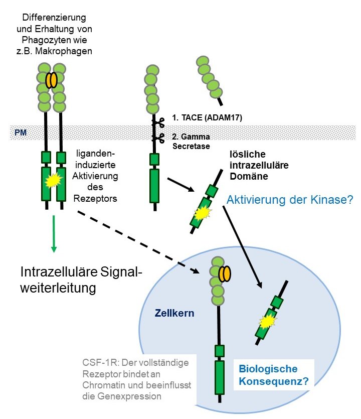
Mutated or aberrantly expressed receptor tyrosine kinases can act as oncogenic drivers in many types of cancer. Signals triggered by the appropriate ligand are already well characterized. However, proteolytic cleavage events may release the intracellular kinase domain of several receptor tyrosine kinases. In addition, both complete receptors and the intracellular domains have been found in the cell nucleus. Whether and how signals via these pathways differ from classical ligand-induced signaling is poorly understood. An example of such signals is the macrophage colony stimulating-factor receptor, CSF-1R, which is normally expressed in phagocytes, but is also aberrantly expressed in epithelial cancer cells. We have shown that the receptor could contribute to survival of epithelial cancer cells under treatment with an anticancer drug. At the moment we are investigating the mechanism by which the intracellular domain enters the nucleus and the potential biological role of its nuclear kinase activity.